

")
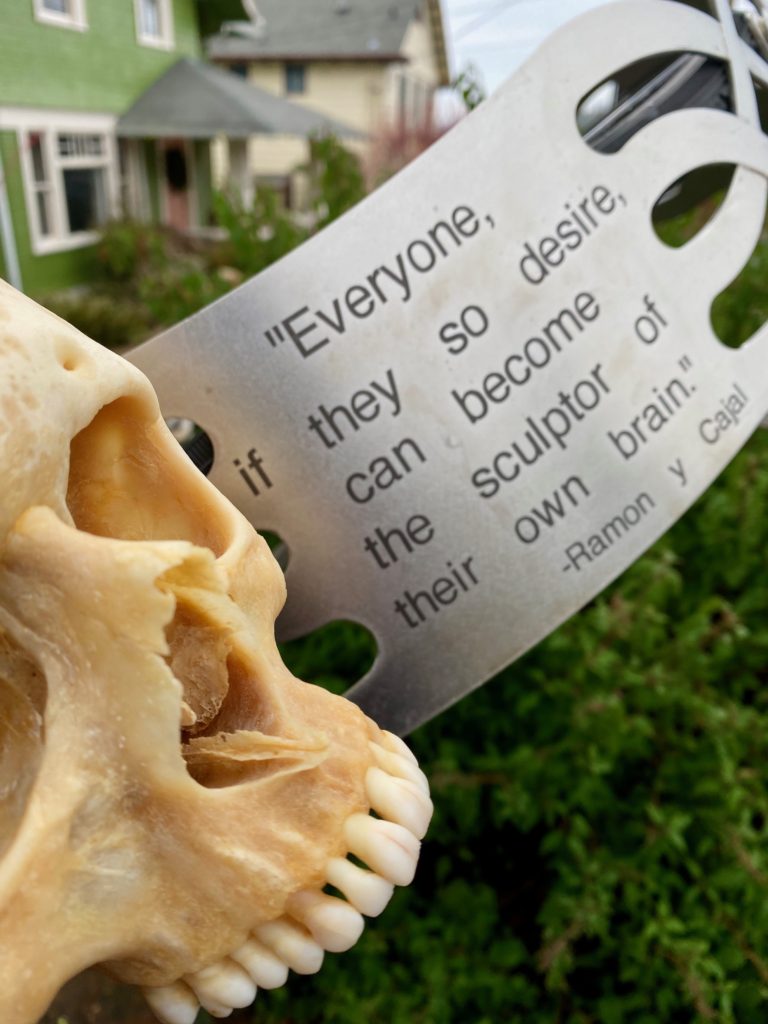
“Life’s true face is the skull.”
― Nikos Kazantzakis
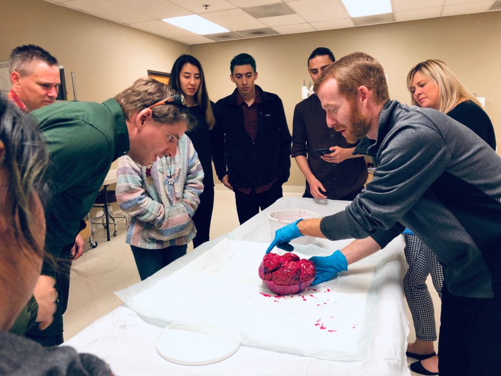
We LOVE teaching anatomy and neuroanatomy!
Few disciplines connect us more viscerally with the extraordinarily physical nature of who we all are.
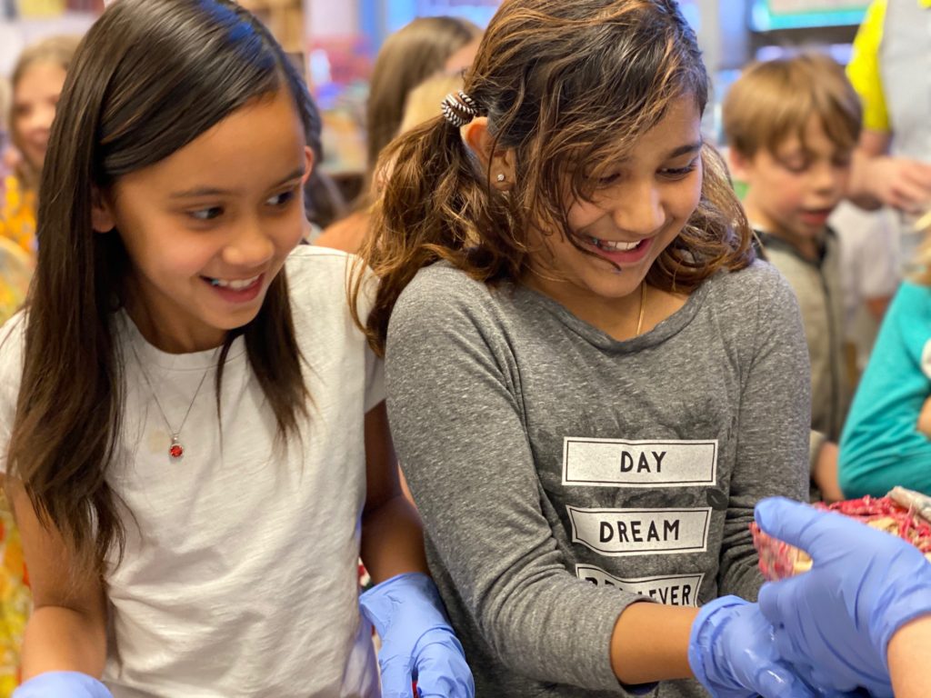
For so many – including artists, educators, researchers, undergraduates, K-12 students, members of the public – the opportunity to directly examine the biological underpinnings of our perceptions, thoughts, ideas, dreams, memories and behavior is astonishingly compelling.
“I profess to learn and to teach anatomy not from books but from dissections, not from the tenets of Philosophers but from the fabric of Nature.”
― William Harvey
Yet such valuable, direct educational experiences weren’t always accessible.
“…in Europe during the Middle Ages, the development of rational thought and investigation was paralysed by the church authorities…During this period, human dissection was considered to be blasphemous…”
― Sanjib Kumar Ghosh
LEARN MORE: Human cadaveric dissection: a historical account from ancient Greece to the modern era
LEARN MORE: The Science of Anatomy
We’d like to thank BioGift Anatomical for their fantastic support of paywall-free, all volunteer arts-integrated neuroscience outreach through NW Noggin.
LEARN MORE: A BioGift of Brains
LEARN MORE: BioGifting brains
LEARN MORE: Adventure A-Head
LEARN MORE: Securing Brains @ BioGift
We’re informed and inspired by the stories we’re told, and the questions asked, research shared, and the art created because of connections made with communities across the country.

Specimens help make this possible!

“Inquiry is fatal to certainty.”
― Will Durant
LEARN MORE: The birth and evolution of neuroscience through cadaveric dissection
LEARN MORE: The importance of scientific collecting and natural history museums for comparative neuroanatomy
LEARN MORE: Retracing the etymology of terms in neuroanatomy
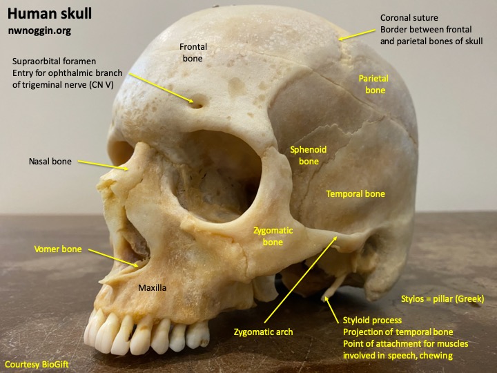
Thanks to the generosity of BioGift, we can now share a genuine human skull, from an 80 year old donor in the Pacific Northwest who’d had brain surgery while alive.
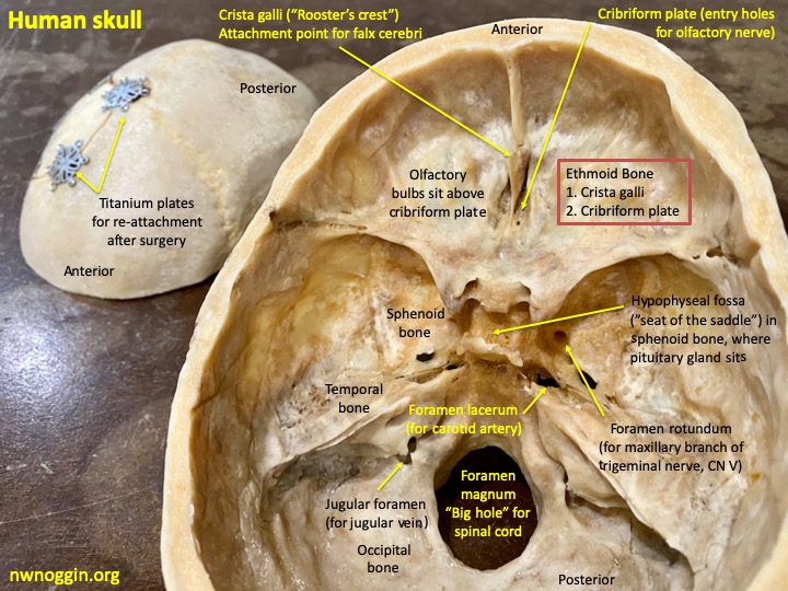

“The skull is a complex component of the skeletal system whose main function is to protect the brain. It is comprised of 22 bones, eight of which form the neurocranium and are connected by synarthrodial joints called sutures.”
― Elizabeth M Lillie et al
LEARN MORE: Anatomy, Head and Neck, Coronal Suture
LEARN MORE: Estimation of skull table thickness with clinical CT and validation with microCT
LEARN MORE: Mechanism of skull suture maintenance and interdigitation

Our NW Noggin participants, along with students we’ve volunteered with at area high schools, have been privileged to witness real brain surgeries thanks to the exceptional Providence Brain Watch program.

LEARN MORE: What is brain surgery like?
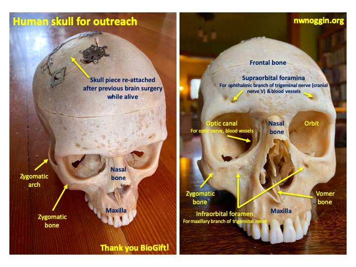
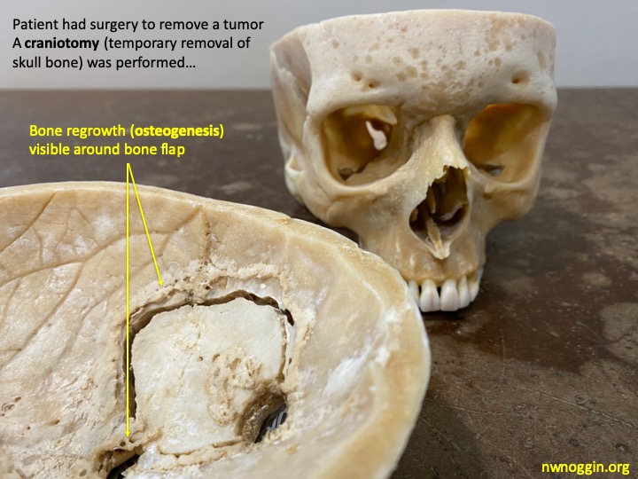
But it’s also incredible and informative to see, touch and examine the actual titanium straps used to cover the drill burr holes and re-connect the bone piece after a surgical craniotomy, up close in an individual’s skull.


For example, those two little holes (above), we know from past surgical viewing, are where the surgeon stitched the leathery meningeal dura mater to the bone (“dural tenting”), to prevent blood and leaking cerebrospinal fluid (CSF) from pooling above.
LEARN MORE: Craniotomy through the ages
LEARN MORE: Awake craniotomy for tumor resection
LEARN MORE: The cranial dura mater: a review of its history, embryology, and anatomy
LEARN MORE: Necessity of dural tenting sutures in modern neurosurgery: protocol for a systematic review
LEARN MORE: Galeal Tack-Up Sutures to Prevent Subgaleal Cerebrospinal Fluid Collection
Learning from direct observation and handling can teach you so much. For example, the slender styloid processes, where muscles critical for speech and chewing attach, appear remarkably fragile (thankfully your own are protected by muscles and surrounding bones!). And portions of the temporal bone – and eye orbits – are surprisingly thin!

LEARN MORE: Anatomy, Head and Neck, Styloid Process
LEARN MORE: Orbital Anatomy for the Surgeon
LEARN MORE: Anatomy, Head and Neck, Orbit Bones
LEARN MORE: Orbital Floor Fracture
LEARN MORE: Calvarial thickness and its relation to cranial bone harvest
People, of course, have explored how our brains and bodies are structured for millennia – and have done so all over the world.
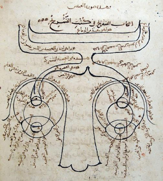
“The seeker after the truth is not one who studies the writings of the ancients and, following his natural disposition, puts his trust in them, but rather the one who suspects his faith in them and questions what he gathers from them, the one who submits to argument and demonstration, and not to the sayings of a human being whose nature is fraught with all kinds of imperfection and deficiency.”
― Ibn al-Haytham
LEARN MORE: Ibn Al-Haytham: Father of Modern Optics
Thanks to exceptional community collaborators, we look forward to many more fascinating questions, arguments, demonstrations, observations, experiments and art!
LEARN MORE: NW Noggin Collaborators