

")
Post by Michael Deveney, NW Noggin volunteer and Portland State University teaching assistant for the Advanced Neurophysiological Psychology course (Winter term 2019)
“Surgeons must be very careful
When they take the knife!
Underneath their fine incisions
Stirs the Culprit – Life!”
–Emily Dickinson
Michael joined fellow NW Noggin volunteers, along with students and teachers from Fort Vancouver High School last week to attend Providence Brain Watch, an innovative educational outreach program that introduces young people to healthcare careers – and neuroscience – by inviting them to watch a brain surgery! Here is Michael’s description of her day in the hospital…

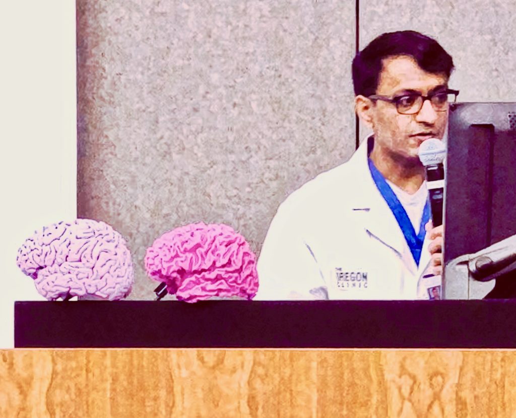
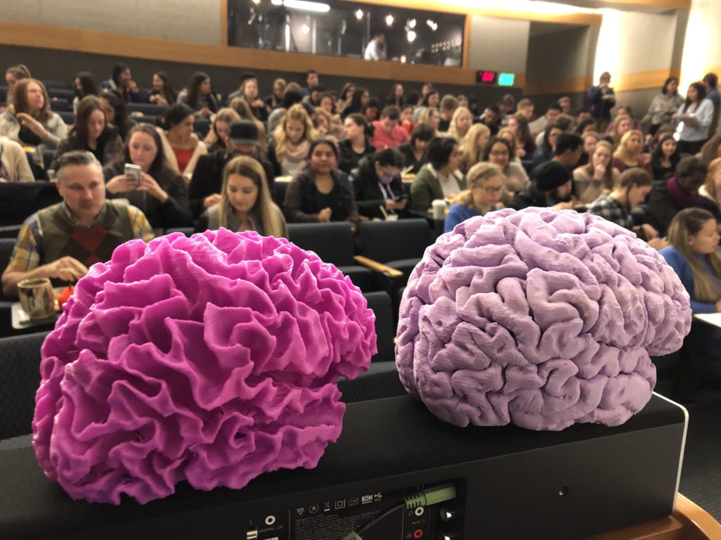
Michael at the OHSU OMSI Brain Fair in March 2018
LEARN MORE: Putting the brains in Brain Fair!
It’s 6 am, and this Facebook post simply NEEDS to be written:
“Good Morning! What are your plans today? I’m just starting my day watching a brain surgery, no big deal.”
That’s how the morning of January 16th started for me, several of my Portland State University classmates, and a large group of high school students from Fort Vancouver! I was graciously invited to partake in this amazing experience by NW Noggin, thanks to the Brain Watch program at Providence St. Vincent Hospital. When I received the invite, I didn’t hesitate. I knew I absolutely needed to be there to learn and discover more about neuroscience, as it may be the only human brain surgery I’ll ever see.
We had a detailed introduction from the neurosurgeon, Dr. Vivek Deshmukh, who described the procedure and his preparation techniques. He explained that he would be removing a vascular mass referred to as a cerebral cavernous malformation (CCM) in a 56 year old male who apparently had over 100 of these in his brain and spinal cord!
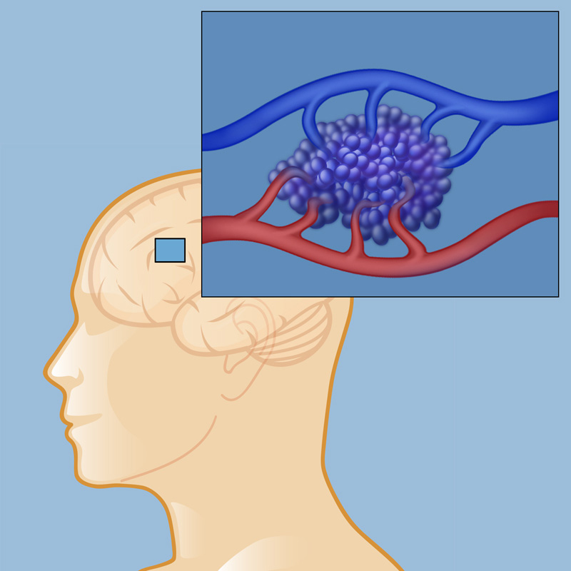
It looks like a mulberry! IMAGE SOURCE: Cavernous Malformations
LEARN MORE: Cerebral Cavernous Malformation Information Page
The patient had lived with these CCMs for his entire life, and most cause no symptoms unless they leak (hemorrhage) or start growing to the point of inhibiting normal brain and body function – which is exactly what this particular one was doing. The malformation was located within the cerebellum in the patient’s right hemisphere, and he was starting to have balance issues, which makes sense considering that our cerebellum is critical for motor control.
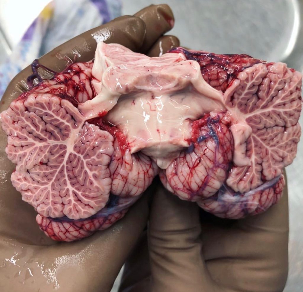
Extraordinary image of the cerebellum by 123Anatomy_human321
After the procedure was explained, several other hospital staff came in to present on their duties while the surgeon scrubbed in to operate. There was a nurse practitioner, a physical therapist, and an MRI technician. Once the surgeon was ready, we were presented with not only the live feed recording, but also a real-time MRI. The surgeon had an earpiece in to answer questions along the way, and a surgical technician was in the room with us to explain the doctor’s every move.
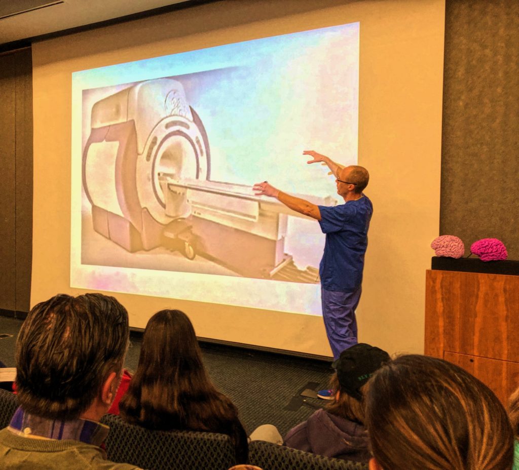
LEARN MORE: The surgical advantages of intraoperative MRI
LEARN MORE: Nurse Anesthetists, Nurse Midwives, and Nurse Practitioners
LEARN MORE: Physical Therapists
LEARN MORE: Radiologic and MRI Technologists
The scalpel raised… We all watched in awe, holding our breath as Dr. Deshmukh made the first slice into the patient’s head, cutting through several centimeters of muscle. Once he got to the skull, a drill was used to cut a bone flap large enough to facilitate the removal of the mass. When the surgeon got close to the dura matter, the tough outer layer of the meninges which protect the brain, he used a 60,000 RPM drill with a footplate to prevent damage to essential blood vessels and the dura itself.
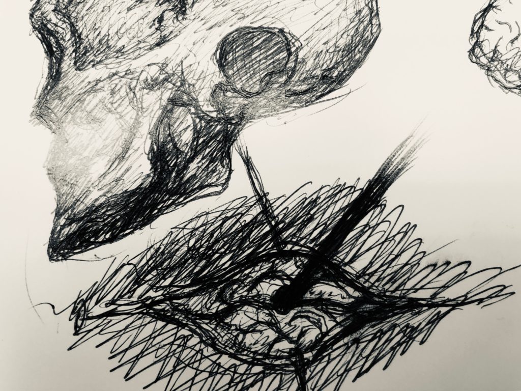
Sketch by NW Noggin Arts Coordinator Jeff Leake
When the bone flap was removed, the surgeon cut into the cerebellum until he could see the malformation, and slowly worked around the mass to remove it without causing any further damage. We learned that a common concern are dystrophic calcifications, rock-like calcium formations (“brain stones!”) that make it more difficult to remove the mass. This patient had many, but you would never know it was an issue watching the surgeon cut around with such grace and ease.
LEARN MORE: Brain stones revisited—between a rock and a hard place
Finally, the big moment came when he had freed the mass and was ready to remove it. As he did, audible exhales of relief were heard across the room, and he showed us a 2.4 centimeter blob of tissue. He quickly stitched the patient closed, working seamlessly…

Dr. Deshmukh then returned to the room to answer more questions. You would never know he had just operated, as he was calm, cool, and collected. Our high school students had so many excellent questions, and were clearly inspired by this exposure to neurosurgery and clinical careers.

This was truly one of the most amazing events I’ve experienced (I even managed to get a selfie with the surgeon!). Thank you NW Noggin and Providence a thousand times over for this truly once in a lifetime opportunity.
LEARN MORE: Noggin @ BRAIN WATCH!
LEARN MORE: What is brain surgery like?