

")
Post written by Kerry Barahona, undergraduate in Psychology and Interdisciplinary Neuroscience at Portland State University. Kerry has interned in Dr. Laura Villasana‘s lab at Legacy Research Institute.
If you asked me fifteen, ten, or even five years ago, I never would have said that I saw myself in research or interested in neuroscience.
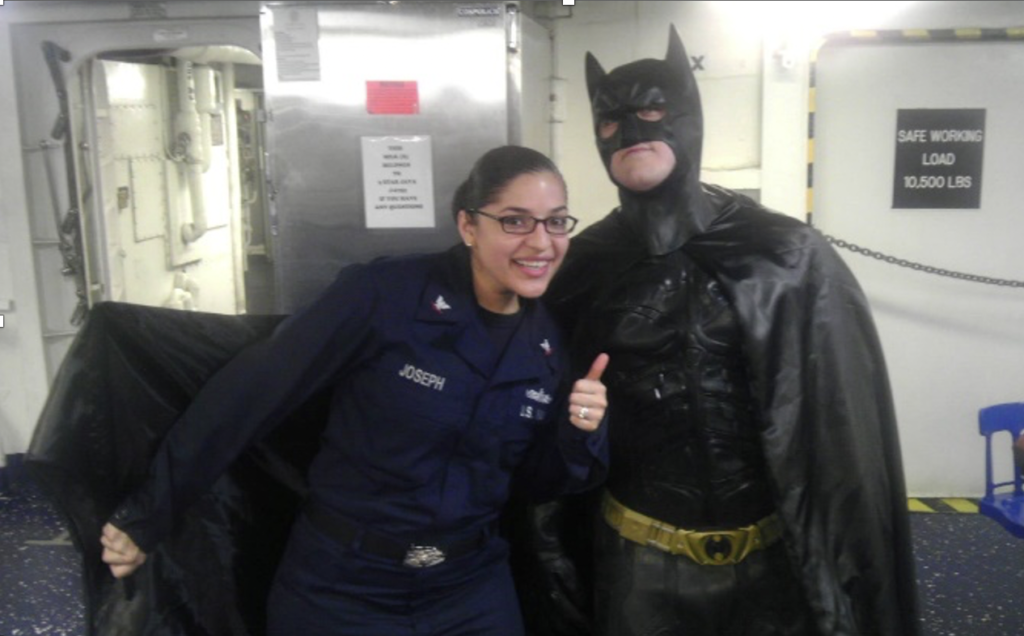
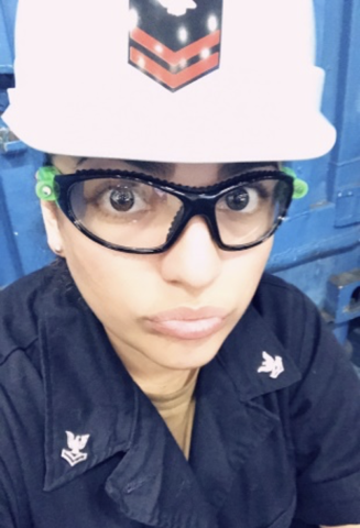
I joined the Navy at 17 years young, needing parental consent and ready to make the biggest sacrifice to get the economic means to go to school. Little did I know that I would absolutely love being a machinists’ mate (a “mechanic” in simpler terms), traveling, and being a sailor.
LEARN MORE: Machinist’s Mate (U.S. Navy)
LEARN MORE: Does the Military Pay for College?
LEARN MORE: Borrow or Serve? An Economic Analysis of Options for Financing Medical Education
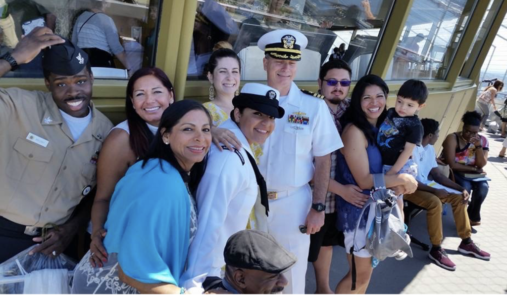
When I reached the end of my Navy contract, after over a decade of service, I went back to school to get my undergraduate degree in pre-medicine. In pursuit of that goal, I changed my major from biology to psychology and ended up taking an introductory neuroscience class at Portland State University…and boy did that change everything.
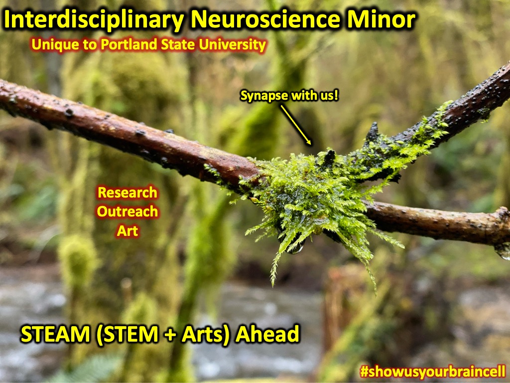
PSU recently added an interdisciplinary minor in neuroscience, and it required four credits of research. Enter Legacy Research Institute, Dr. Laura Villasana, a summer paid internship, and my first job in a neuroscience lab. Even more kismet? The research I am engaged in can be applied directly to military service members (I’d say more, but they might take me out ;p if you catch my drift).
This type of research is important to me because it has affected me both professionally with my comrades, and personally, with my brother-in-law.
LEARN MORE: Interdisciplinary Neuroscience Minor @ Portland State University
LEARN MORE: A Legacy of Links
Traumatic Brain Injury
I was very excited to contribute to work in Dr. Laura Villasana’s lab at Legacy. The research I became involved with examined the impact of a special diet on individuals with traumatic brain injuries (TBIs).
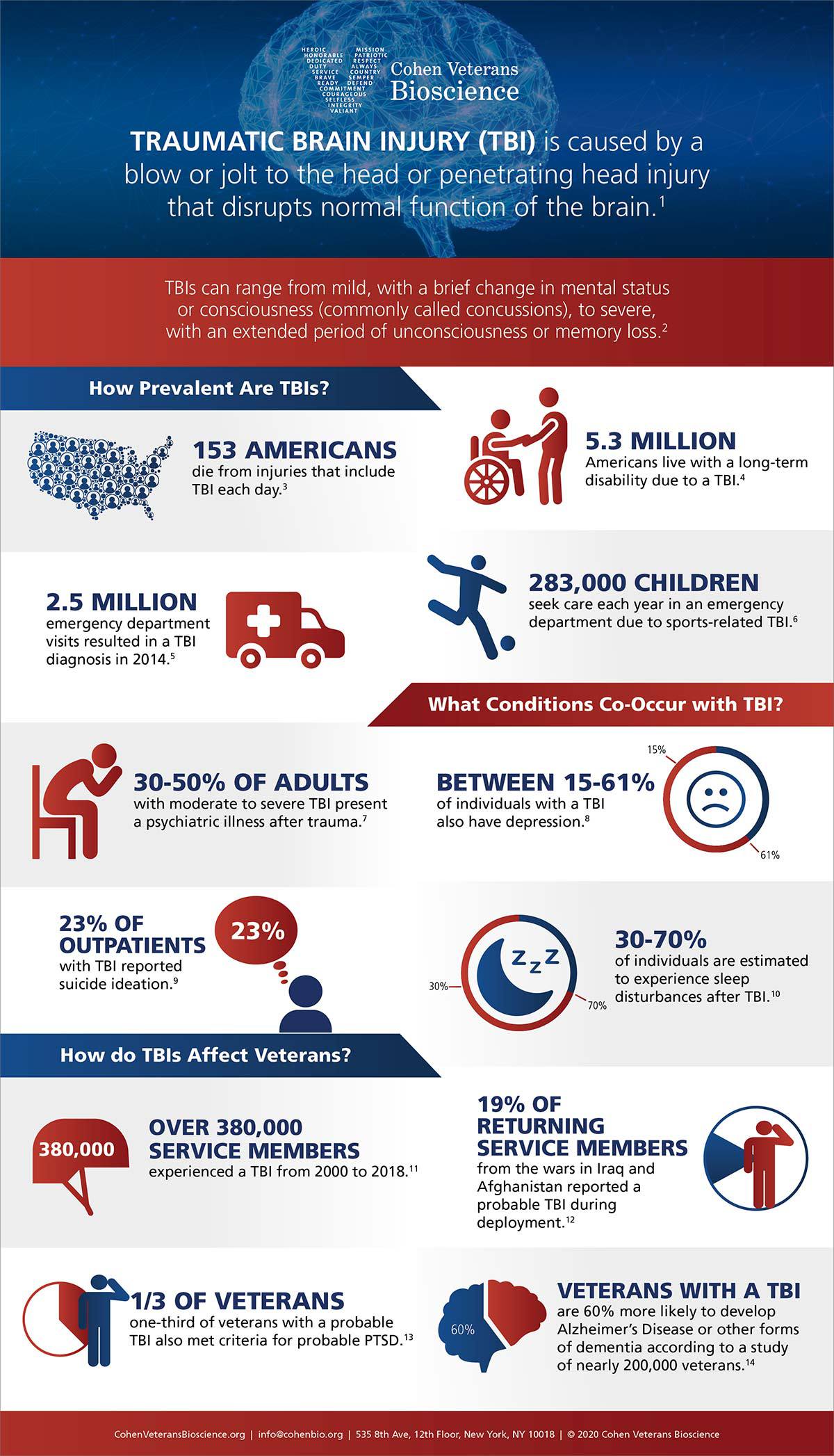
TBIs are unfortunately common, and usually result from a hit or jolt to the head or body.
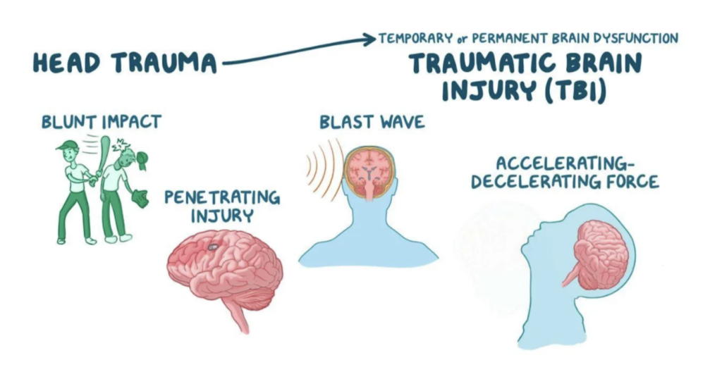
A minor TBI affects your brain cells temporarily, but more serious or severe TBIs can lead to bleeding, bruising, torn tissue, and enduring physical damage to the brain. These injuries can result in long-term symptoms and complications, or even death. TBIs can cause sensory issues (e.g., difficulty processing what you see, or hear) as well as cognitive and behavioral issues, including memory loss, mood swings, confusion, anxiety and depression. Many veterans are still recovering from TBIs, are are considered a “population of special concern.”
LEARN MORE: Traumatic Brain Injury: Hope Through Research
LEARN MORE: Traumatic Brain Injury (NIH)
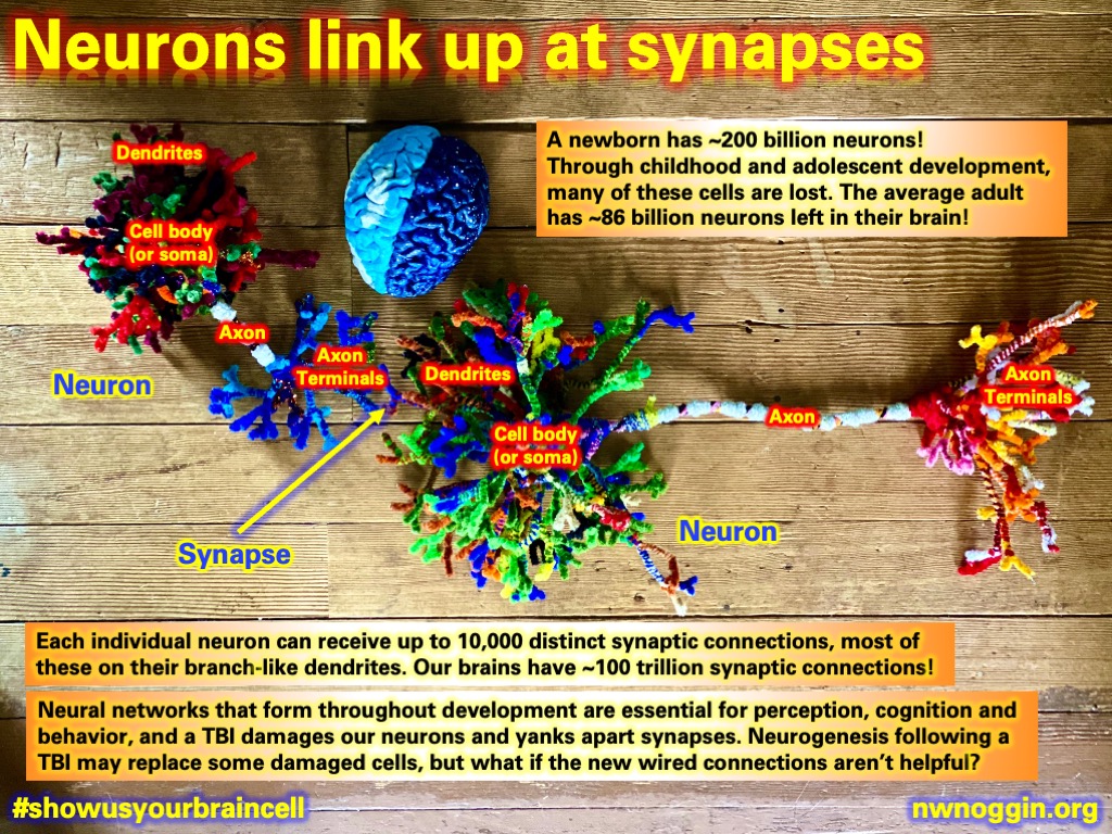
When a TBI occurs, there is a sudden surge in neurogenesis, or the process by which new neurons are formed in the brain. Some researchers suggest that neurogenesis can be beneficial. There are some growth factors and drugs shown to enhance neurogenesis and improve functional recovery.
LEARN MORE: ADULT NEUROGENESIS IN HUMANS: A Review
LEARN MORE: Factors that influence adult neurogenesis as potential therapy
LEARN MORE: The role of adult hippocampal neurogenesis in brain health and disease
Can a Nicotinamide Riboside (NR) Diet Help with TBI?
The NR diet is one that supports healthy aging, cellular production, brain function, liver health, and cellular metabolism (essentially cell support in cellular processes).

LEARN MORE: Nicotinamide Riboside—The Current State of Research and Therapeutic Uses
LEARN MORE: The Influence of Nicotinamide on Health and Disease in the Central Nervous System
Nicotinamide riboside (NR) is an antioxidant that helps restore adequate levels of nicotinamide adenine dinucleotide (NAD), an important enzyme that is needed for cellular and mitochondrial functions (which are interrupted after TBIs). Essentially, NAD immerses brain cells in nutrients, resulting in improved brain health and function. A decline in neurogenesis is caused at least partly by a decline in NAD, which leads to an interruption or stall in mitochondrial functions. When brain cells known as radial glia cells decrease in number following injury, NAD decreases as well. The idea is that with NAD increase, glial proliferation will increase along with neurogenesis from neuronal stem cells in the brain.
LEARN MORE: Impact of Traumatic Brain Injury on Neurogenesis
LEARN MORE: Neurologic impairment following closed head injury predicts post-traumatic neurogenesis
This research project is examining whether the NR diet in mice impacts neurogenesis after a TBI, as well as how TBI severity is influenced by the diet. I was struck by how we don’t really know whether neurogenesis is always beneficial or not. I guess the answer that we have so far is “it depends.“
Dr. Villasana is researching this, and she suspects that there could be some negative consequences with neurogenesis following TBIs because the process might potentially establish some “bad wiring.”
I was very excited to analyze and gather data for this project, especially since military members suffer from TBIs at a higher rate than most professions.
When I was given a list of projects to consider joining, I was not told that this research had a military association because my principal investigator (PI) didn’t want to bias my decision. We were both very happy when I selected this project, and even more so when I realized that this could be a possible treatment for fellow service members suffering and recovering from TBIs.
LEARN MORE: Military traumatic brain injury: a challenge straddling neurology and psychiatry
LEARN MORE: Incidence of Traumatic Brain Injury in the U.S. Military, 2010-2014
LEARN MORE: Military-related traumatic brain injury and neurodegeneration
Expectations vs. Reality
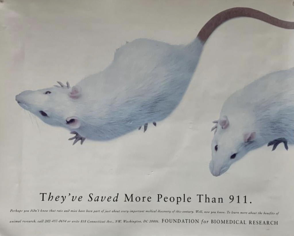
When entering the world of research, I thought it was mostly going to be data and analysis, and while there is a lot of that, there is also a lot of hands-on things that I didn’t expect. First and foremost, I work with mice. I didn’t realize how much training was required to handle mice (and I mean LOTS).
I couldn’t really take pictures of the mice or their living quarters (it’s frowned upon for many reasons), but I did handle them when we needed to sequence their genomes. Genome sequencing is the process in which the order of the genome, or DNA fingerprint, is determined or read.
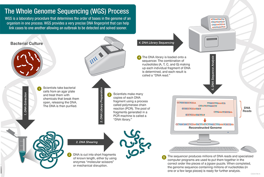
LEARN MORE: What is whole genome sequencing (WGS)?
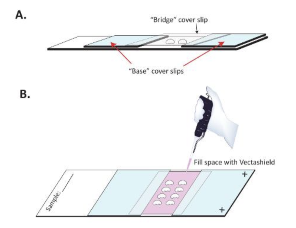
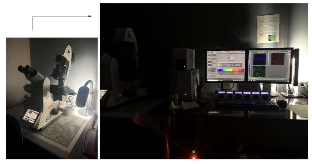
Second, I didn’t realize that I would spend so much time sitting in a dark room imaging mounted slides. Mounting the slides is also not as easy as it looks, you must ensure that the brain slices are flat and wrinkle and bubble free. They are then left to dry in a slide storage box laying flat so that the mounting media can be added and then covered with a single glass cover. Everyone has their own technique, but essentially you use delicate paint brushes to mount your subject, then add the mounting media (usually VectaShield), then cover it and seal the edges with clear nail polish and let them dry.
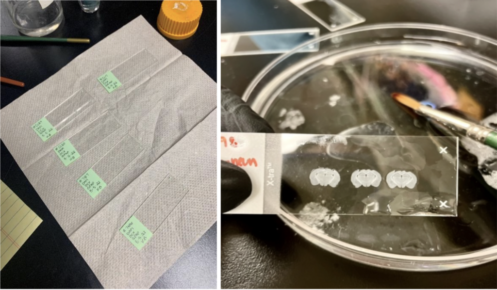
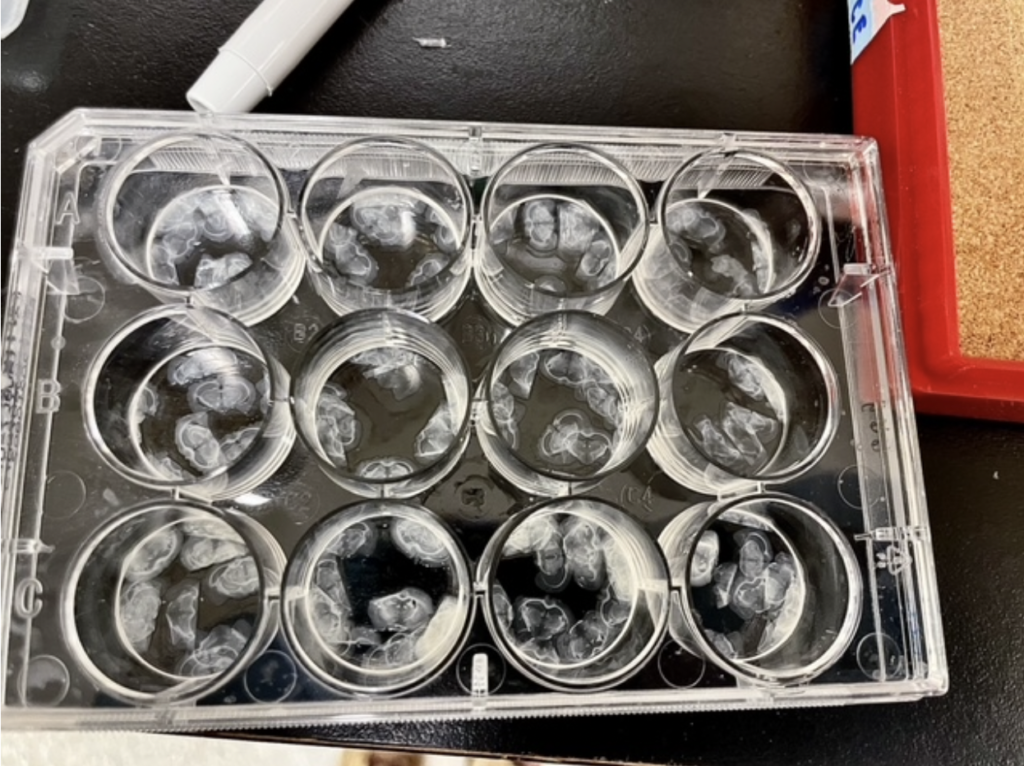
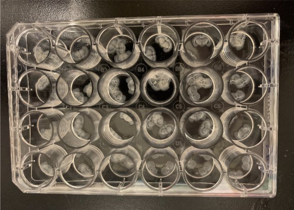
There is also a lot of storing. Moving brain slices from wells into centrifuge tubes to prevent loss of materials (because it can mold if not stored correctly, but I mean come on, aren’t they so pretty?!).
I also didn’t expect to love research!
I worried that I was going to hate it.
I thought it might be boring and tedious and that my days were going to drag on and on while at the lab.
I was so glad to be proven wrong. I love my job!
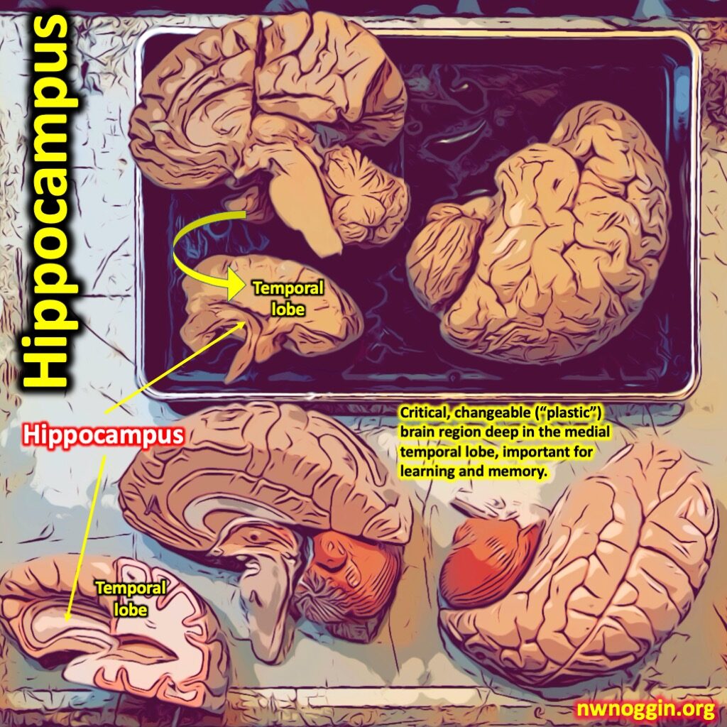
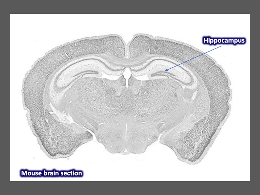
I love that I know how to recognize the beginning of the hippocampus, the location of neurogenesis.
LEARN MORE: A closer look at the hippocampus and memory
LEARN MORE: How the hippocampus contributes to memory, navigation and cognition
LEARN MORE: The role of adult hippocampal neurogenesis in brain health and disease
I’m also thrilled that I’ve been trained to use a cryostat machine that cuts tissue at low temperatures (we keep ours at -20°C) as small as 4 μm (microns). Most tissues are cut between 6-15 microns in thickness, sometimes as thin as 4 microns, whereas our tissue is cut much thicker, above 50 microns.
LEARN MORE: What Is a Cryostat & How Does It Work?
The Beauty of the Brain
I also didn’t think that I would love the analysis portion of research, or that I would spontaneously say “it’s so pretty” or “beautiful” multiple times a day! But it is so pretty and beautiful – just look!
LEARN MORE: A Beautiful Science
LEARN MORE: The Art of the BRAIN
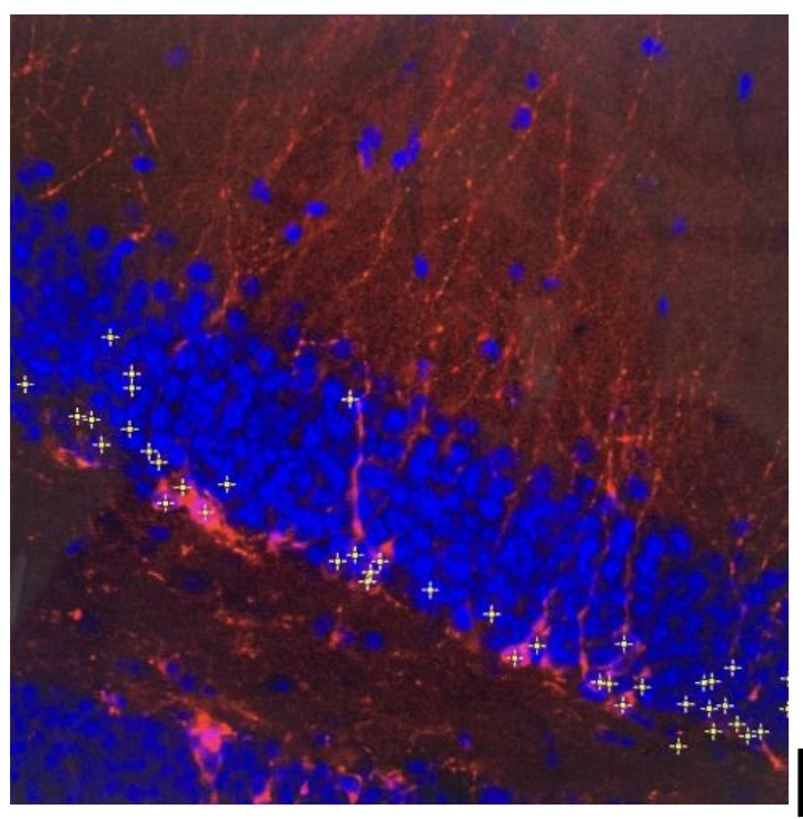
The tissue we cut and analyze is thicker so that we can use z-stacks to determine the number of cells in that subject. This is viewed via a confocal microscope, using x- and y-axes that remain the same, but the z-axis is different, like traveling through the tissue. I think that’s when it’s most beautiful because you can see the neurons and axons so clearly.

I’m indebted to Dr. Laura Villasana and Legacy Research Institute for welcoming me into the lab! And if you feel like I did before I began this endeavor (like research was the last thing you ever wanted to do) then I suggest that you give it a try. You just might love it too.