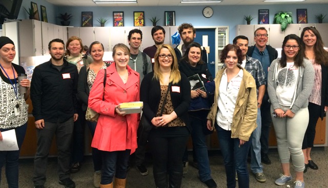
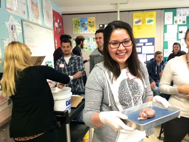
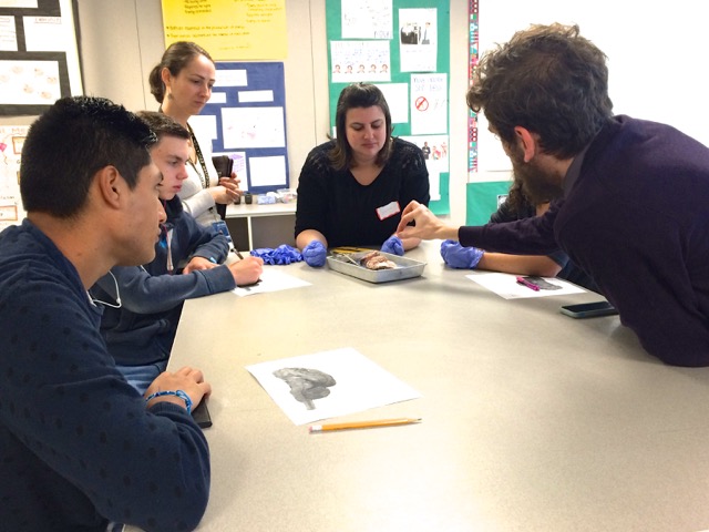
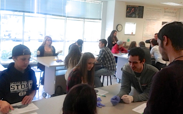
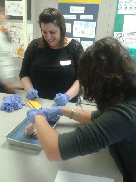
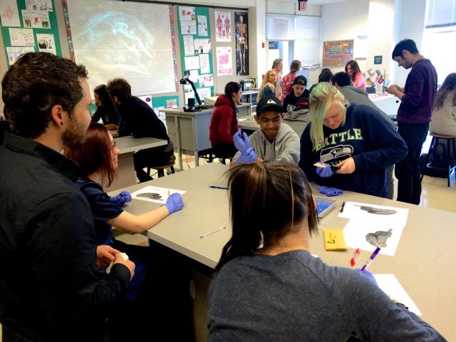
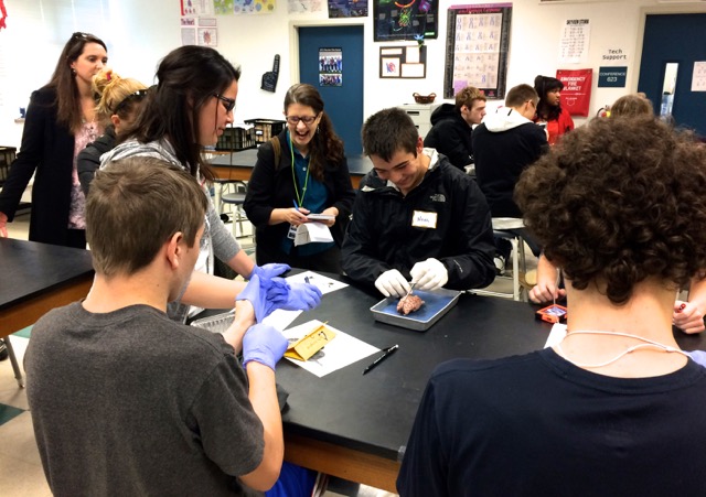
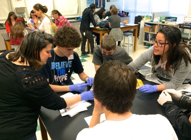
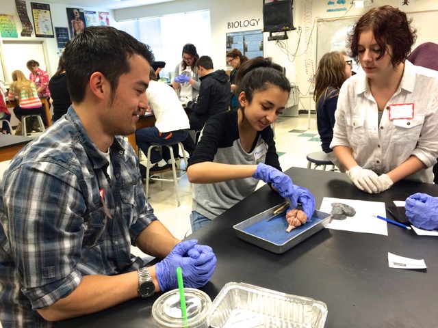
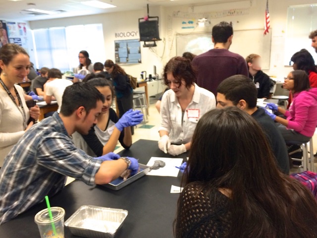
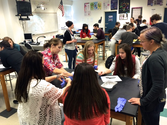
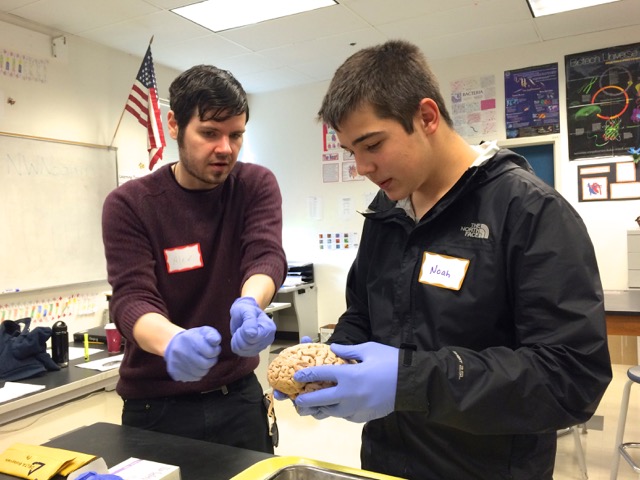
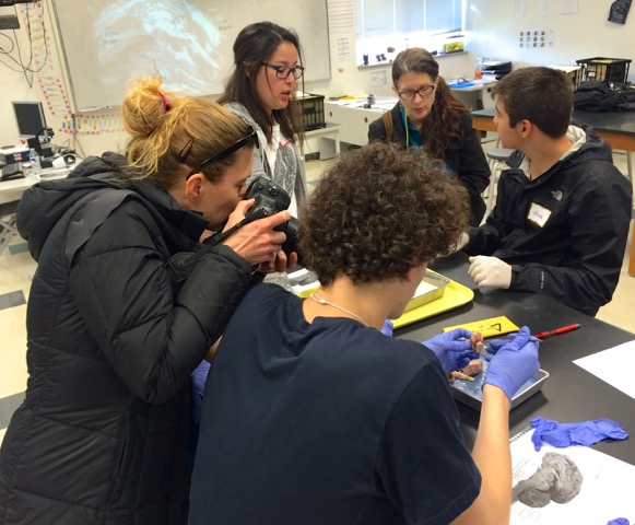
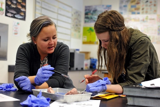
A healthy crowd of committed undergraduate volunteers gathered early this morning at Skyview High, home of the Storm, for our fourth and final day in Biology classrooms…
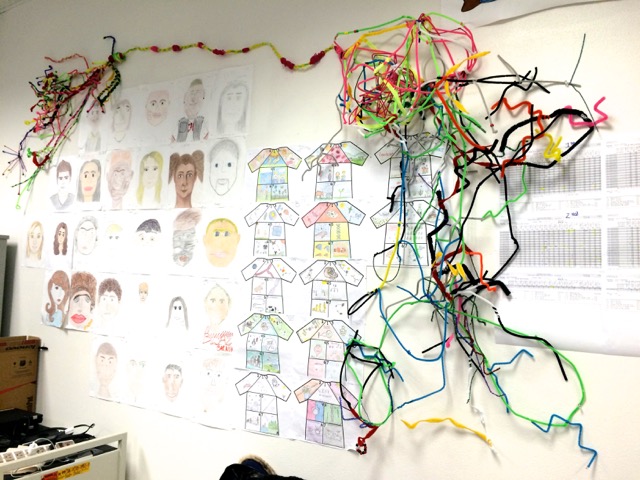
We are going to miss our students, whom we’ve gotten to know over the past month, through our ongoing series of Thursday visits. Art activities, short introductory lectures, and direct, hands on examination of real human and animal cerebrums have done a lot to engage them in learning about their own brains and behavior…
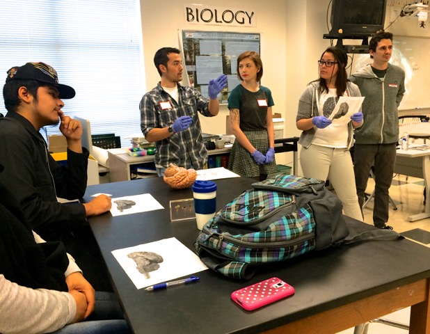
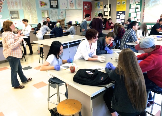
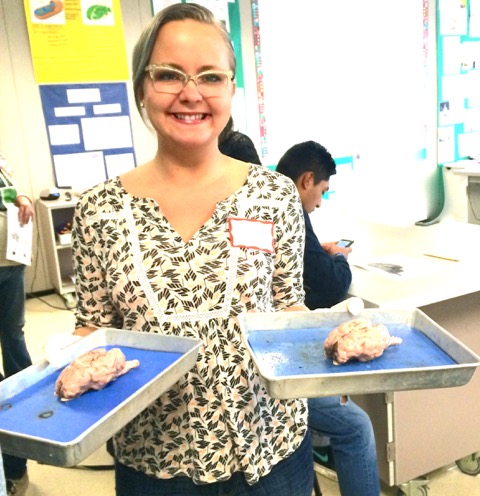
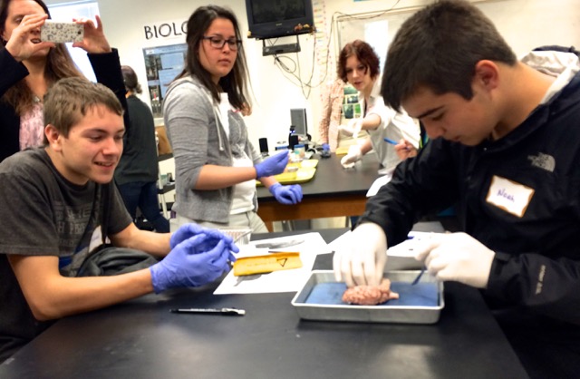
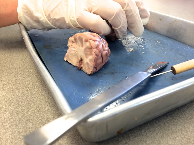
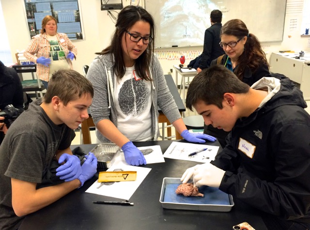
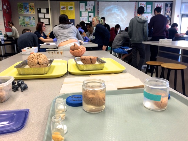
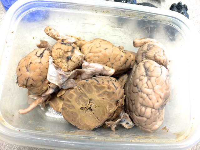
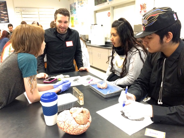
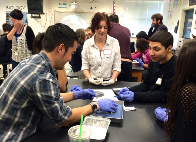
According to Biology teacher Angela Fojtik, attendance on Thursdays has never been better! And today was no exception, as we brought along a bucket of sheep brains, dissection trays, and several sharp scalpels to see what’s under the hood…
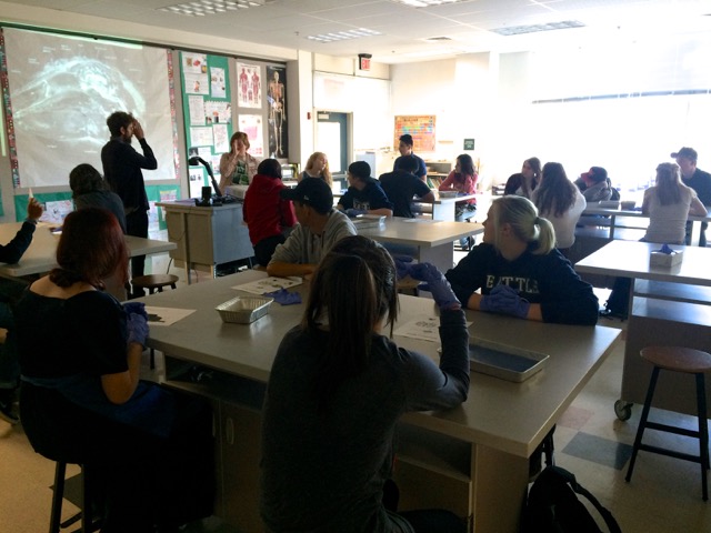
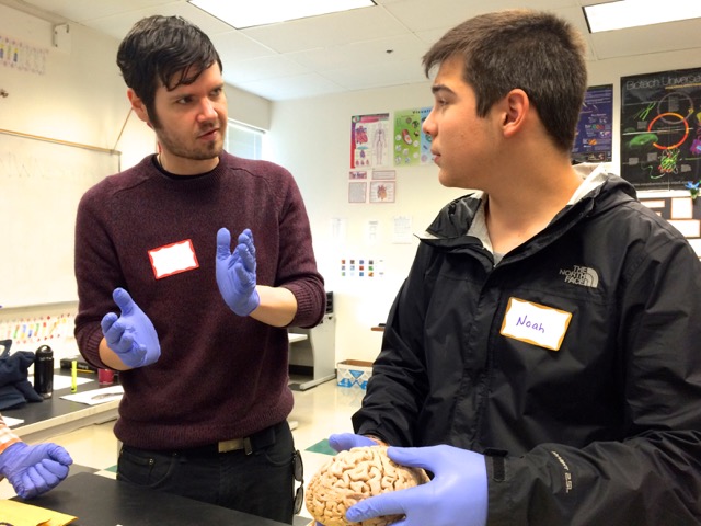
Our accomplished Noggin volunteers led off each class, with Michael Miller, Angela Johnson, Nathan Allen, Angela Gonzales (a former Skyview student), Karlo Valle, Elizabeth Olson, Grace Hoinowski and Elias Shaw taking charge…
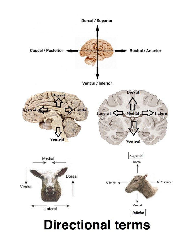
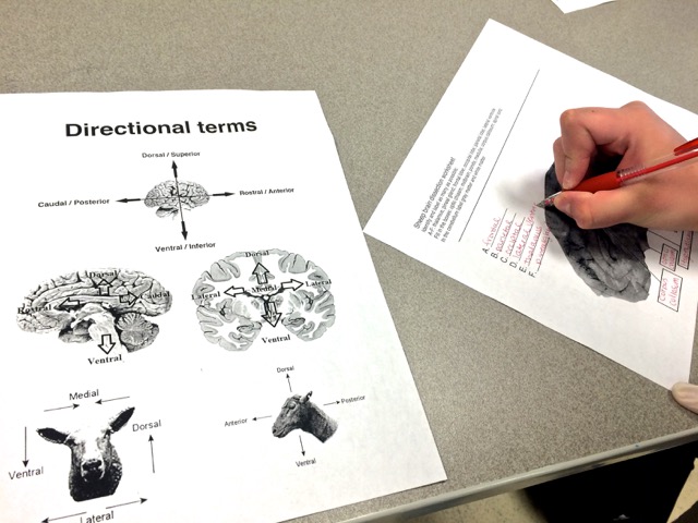
They went over directional terms, explaining how neuroanatomists refer to different parts of the nervous system using words like anterior (or, in the cerebrum, rostral), posterior (or caudal, Latin for “tail”), superior (dorsal), and inferior (ventral)…
Directional terms handout; prepared by Angela Johnson
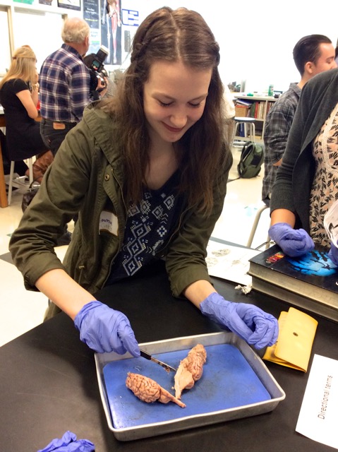
They also described the various cuts you might make through a sheep’s brain, including horizontal (along the horizon), coronal (like a corona around your head), and sagittal…
We also asked our high school students which was larger, a sheep’s brain, or its tongue?
The answer surprised them! Then we started making our cuts…
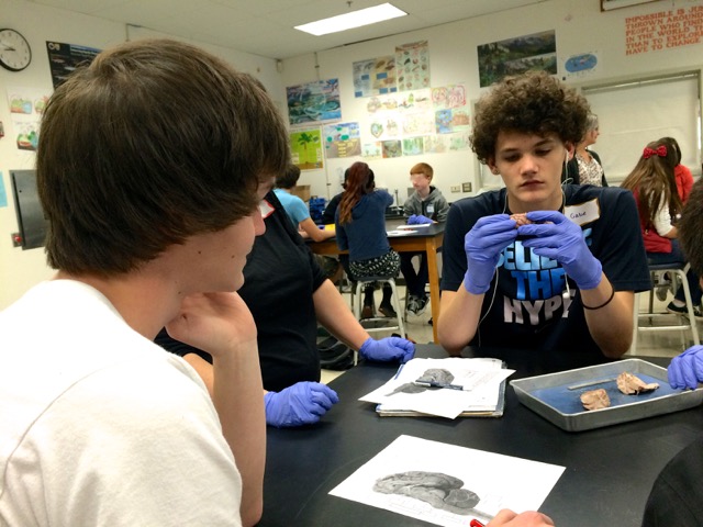
The difference between gray matter (neuron cell bodies, dendrites, and axon terminals; the site of synaptic connections, and chemical communication between cells) and white matter (the axons, or wires, covered by fatty cell membranes from supportive glial cells, carrying electric currents between areas of gray matter) was clearly visible in a coronal slice…
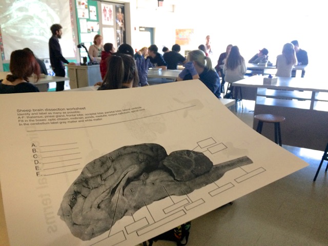
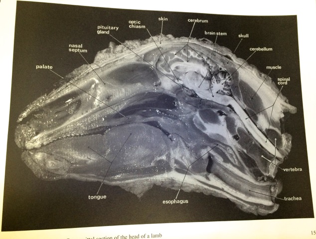
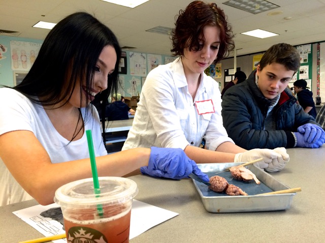
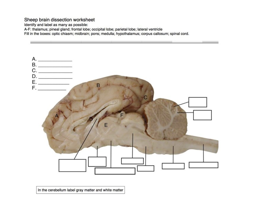
Students identified structures after a mid-sagittal cut, including the cerebellum, medulla, pons, midbrain, corpus callosum, hypothalamus, thalamus, optic chiasm, and pineal gland… They filled out a handout for extra classroom credit
Sheep brain dissection; handout prepared by Angela Johnson
There are few things more compelling and motivating than exploring the (often smelly :)) neural circuitry responsible for cognition, perception, and behavior, and directly comparing the brains of different species…
The small groups, and numerous undergraduates from WSU, and PSU, encouraged questions about the functional role of different brain structures, including the pineal gland. Students were very interested in its night time release of melatonin, and role in circadian rhythms. We discussed how melatonin promotes sleep, and how pineal gland release of this hormone stops in response to light exposure. Light at night – from our cell phones, and tablets – has been linked to decreased melatonin release, and poorer sleep…
This understanding of the role of specific brain networks that respond to environmental stimuli with behaviorally relevant responses offers students real insight into changes they might make to improve their own lives…
A reporter and photographer from The Columbian newspaper joined us during our second period class, and produced the following story…
Skyview students go inside the noggin
During our third classroom visit, we were joined by a representative from Vancouver Public Schools, who published another writeup…
Brains, brains everywhere – and lots of thoughts to think
VPS also produced a video (we are story #2)…
A great series of visits – and a Skyview Storm of brains – that we plan to repeat in other area public schools this spring. Many thanks to Angela Fojtik of Skyview High, and Nina Stemm and Hannah Valenti of GEAR UP, for welcoming us to class!


