

")
Post by Kirby Kahl, undergraduate in Psychology with a minor in Interdisciplinary Neuroscience at Portland State University. Kirby is a research intern in the Moss Lab at Oregon Health & Science University studying the consequences of traumatic brain injuries (TBIs). She is supported by the McNair and URISE Scholarship Programs at PSU.
In the beginning…
I became interested in traumatic brain injuries (TBIs) following my own experience with one in 2022. I’m curious about resiliency and how our brains recover from insults, both mechanistically (what actually happens in the brain after a TBI) and how we’re able to re-gain function.
The Centers for Disease Control reports that about three million people suffer brain injuries each year from car accidents, sports activities and more! Head injuries are highly variable and greatly under-reported. I wanted to add to the body of literature on TBIs and how our brains respond.
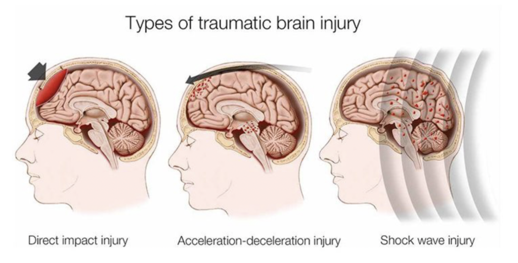
IMAGE: Traumatic Brain Injury
LEARN MORE: Traumatic Brain Injury (TBI)
LEARN MORE: Traumatic Brain Injury & Concussion
LEARN MORE: McNair Scholars Program @ Portland State University
I began my research journey through the McNair Scholarship Program in January 2025 where I developed a research proposal centered on a mouse “model” of TBI.
Scientists often use animals models to implement (“model”) human conditions in order to learn what happens and how to help. Some TBI models are “closed head” models while others are “open head,” which refers to the techniques and surgical invasiveness required by each approach.
Closed head models are considered more closely related to human TBIs, but they are also a lot more variable which can make it hard to get consistent quantifiable results. Open head models, including one called Controlled Cortical Impact (or CCI), are less directly translatable to humans but offer more consistency (i.e., they deliver the same impact every time).
I chose CCI due to its consistency. CCI involves delivering a precise 3mm brain lesion through a 5mm craniotomy (a tiny hole in the cranium, or skull) with a specialized “impactor” on a piston.
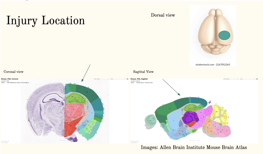
LEARN MORE: Allen Institute Mouse Brain Atlas
LEARN MORE: The Controlled Cortical Impact Model of Experimental Brain Trauma
LEARN MORE: Modeling traumatic brain injury using controlled cortical impact injury
After a TBI, we typically see the growth of new brain cells, which is known as neurogenesis.
Neurogenesis also occurs after other brain insults such as stroke. When many brain cells die off at once, they put out chemical signals that encourage the generation and migration of new cells. As scientists, we are not sure whether neurogenesis is inherently good or bad, but because it occurs after injury, we know that it’s significant and worth understanding.
One of the best ways we have of identifying these adult-born cells following injury is by administering BrdU, a chemical compound that labels new (or proliferating) neurons.
We also call this pulse labeling. Pulse labeling involves injecting BrdU at two different timepoints, usually about four hours apart. BrdU labels cells that are in the s-phase of the cell cycle, when DNA replication occurs, so utilizing a couple of injections allows us to capture a broader range of proliferating cells.
LEARN MORE: BRDU staining
LEARN MORE: An Overview of the Cell Cycle
LEARN MORE: BrdU assay for neurogenesis in rodents
LEARN MORE: Neurogenesis after traumatic brain injury
LEARN MORE: Neurogenesis in Adult Human Brain after Traumatic Brain Injury
LEARN MORE: Versatile strategies for adult neurogenesis: avenues to repair the injured brain
With these experimental methods in mind, I developed a proposal that included pulse labeling new cells with BrdU following a CCI open head model of traumatic brain injury in rodents.
I injected BrdU seven days post-injury with the impactor.
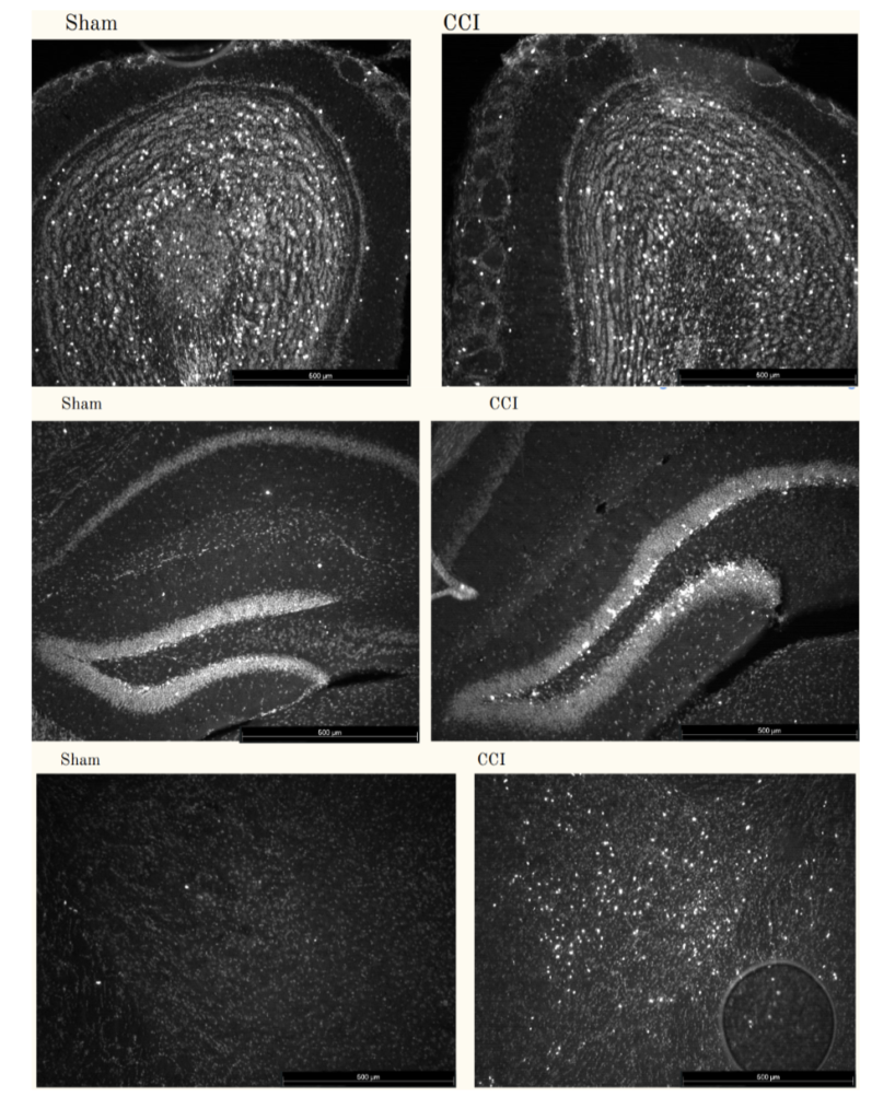
At three weeks post-injury, I “harvested” the brain tissue and preserved it before slicing it into thin sections for further analysis. I then used a form of immunohistochemistry (IHC) which is a mechanism of visualizing antibodies on cells (through fluorescence – these antibodies are tagged with chemicals that glow!) to gather information about them.
When I place my slides containing the thin sections under a microscope, I look for the fluorescent, glowing cells that have been lit up by BrdU and immunohistochemistry. BrdU only labels the cell nuclei, so in the images below, the brightest white lights are representative of new cell bodies.
LEARN MORE: Immunohistochemistry
LEARN MORE: Immunohistochemistry of Brain Tissues
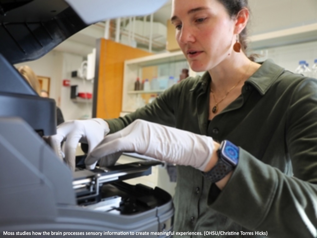
While wrapping up the details on this proposal, I was accepted into the OHSU Neuroscience Graduate Program Summer Internship and was matched with Dr. Elizabeth Moss.
While the summer internship was defunded in the wave of grant cancelations in early 2025, Dr. Moss let me stay to complete my project. I ended up getting cool results, and I also learned how to follow my data, including some surprising data, which we’ll talk about below. That’s a lot of the fun in research!
LEARN MORE: See the alarming extent of NIH and NSF funding cuts in 2025
LEARN MORE: Trump admin axed 383 active clinical trials, dumping over 74K participants
LEARN MORE: The Impact of Federal Funding Cuts on Research, Practice, and Patient Care
LEARN MORE: US science after a year of Trump
Early data
My initial data showed BrdU labeled cells in all sorts of surprising locations like the thalamus, the striatum and more. The images below reveal these new cell nuclei as dots of light, while the graphs show cell counts (how many new cells) by their location in the brain.
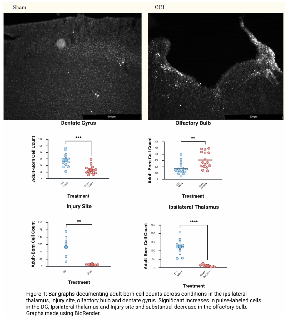
Figures created by Kirby Kahl, for a paper coming later this year
A key piece of information needed to assess and treat brain injuries is how much of the brain and body a TBI can affect. Seeing new brain cells further from the original injury site means that a small injury can have big consequences, even in areas quite far from the injury itself.
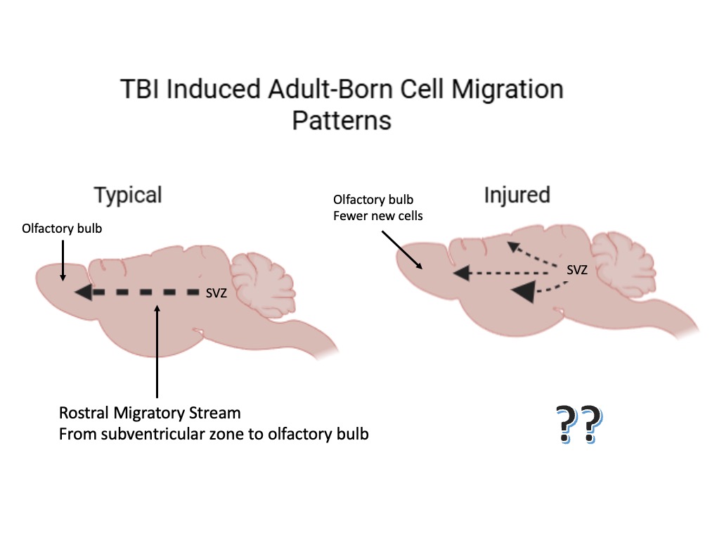
In non-injured brains we also see neurogenesis, or the generation of new neurons.
These new neurons divide from what are called neuronal stem cells in the Subventricular Zone (SVZ), an area around the lateral ventricles. The new cells then migrate from the SVZ to the olfactory bulbs, where they replace older cells. This pattern of neurogenesis and migration continues into adulthood, and the migratory path these cells follow is called the Rostral Migratory Stream (RMS).
LEARN MORE: Neurogenesis in Adult Subventricular Zone
LEARN MORE: Control of neuronal migration through rostral migratory stream in mice
LEARN MORE: Rostral migratory stream neuroblasts turn and change directions in stereotypic patterns
My data also revealed a stark and surprising decrease in new cells in the olfactory bulbs! Given these findings, our hypothesis was that new cells might be diverting from their typical migratory path (the Rostral Migration Stream) and ending up instead in other parts of the brain.
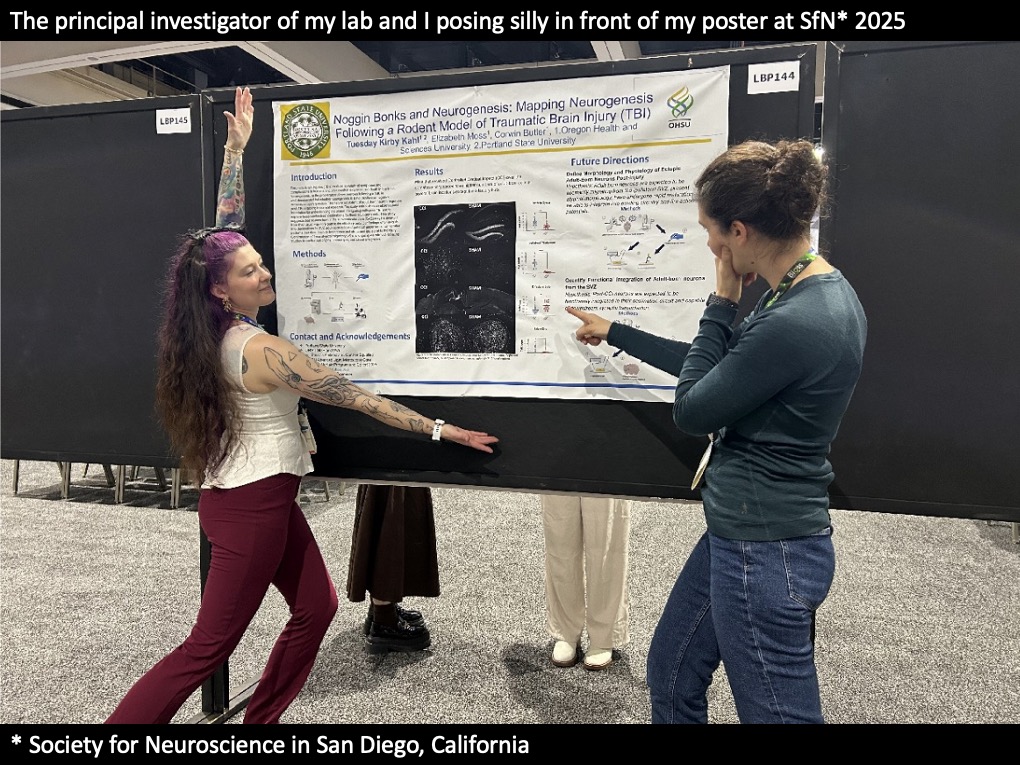
I presented this work at the 2025 Society for Neuroscience (SfN) conference as first author and had a wonderful time learning more about neuroscience and all sorts of cool research!
LEARN MORE: Society for Neuroscience
LEARN MORE: Noggin @ Society for Neuroscience
Right about the time that I attended the conference, I also applied to eight Ph.D. programs in Neuroscience/Neurobiology as I’m immensely excited to continue my research. I also applied for the National Science Foundation Graduate Research Fellowship Program, a grant that supports graduate students. I completed my proposal using the preliminary data from my summer research.
LEARN MORE: What is the NSF Graduate Research Fellowship Program?
Follow the Data
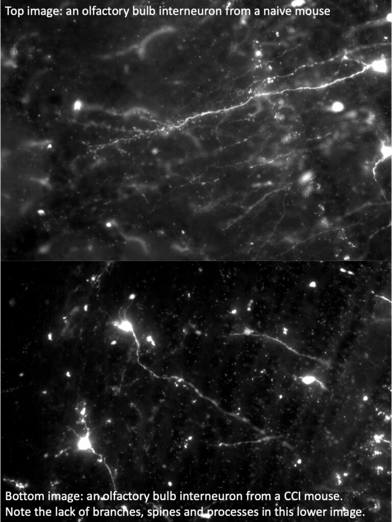
So after conferences and applications I wanted to see if I could label these migrating cells using a different technique. So this time I injected fluorescent lentivirus into the subventricular zone.
Lentiviruses can be used to fluoresce or “light up” dividing cells in a particular location as well as their progeny. We injected lentivirus into the subventricular zone of our mice, then followed with either a naive, sham or CCI surgery and then later processed the tissue using immunohistochemistry. If these cells were in fact diverting along their path to the olfactory bulb, then we should see our virus-labeled cell bodies and processes in other locations, similar to where we saw the labeling with BrdU.
However, we saw pretty minimal viral labeling anywhere other than the olfactory bulbs! So our hypothesis was apparently incorrect, and we still need to understand where all those BrdU-labeled new cells are coming from and how they are ending up in these various locations in the brain.
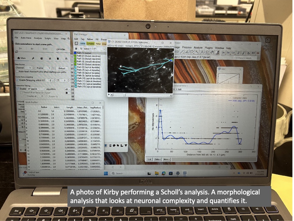
Yet because we used a lentivirus, we actually lit up the entirety of each new neuron (not just cell bodies), including all of its processes like the axons and dendrites. Looking closely we noticed that newly formed cells in the CCI-injured animals that arrive in the olfactory bulb look very abnormal.
What’s Next?
I’ll present this work in April at the Association for Chemoreception Sciences and as my PSU Honors College thesis in June. Getting involved in research has been positively life-changing. It’s an incredible blend of reading, precise hand-eye coordination, creativity and problem-solving that keeps me consistently engaged.
I am excited to continue my work as I begin the OHSU Neuroscience Graduate Program as a Ph.D. student this summer! I’d encourage anyone interested in research to reach out to PIs (Principal Investigators, the “bosses” of a lab) and apply for research programs.
Do not be deterred by rejections. There may be many rejections before an acceptance, but each acceptance then snowballs into the next. Hold fast, read literature, follow your data and stay stoked!