

")
Ben Johnston is finishing his Bachelor of Science in Applied Health and Fitness with a minor in Interdisciplinary Neuroscience at Portland State University. He previously earned an AAS degree in Exercise Science at Portland Community College.
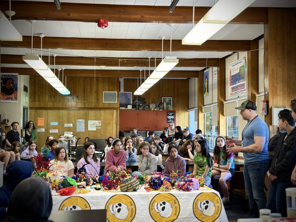
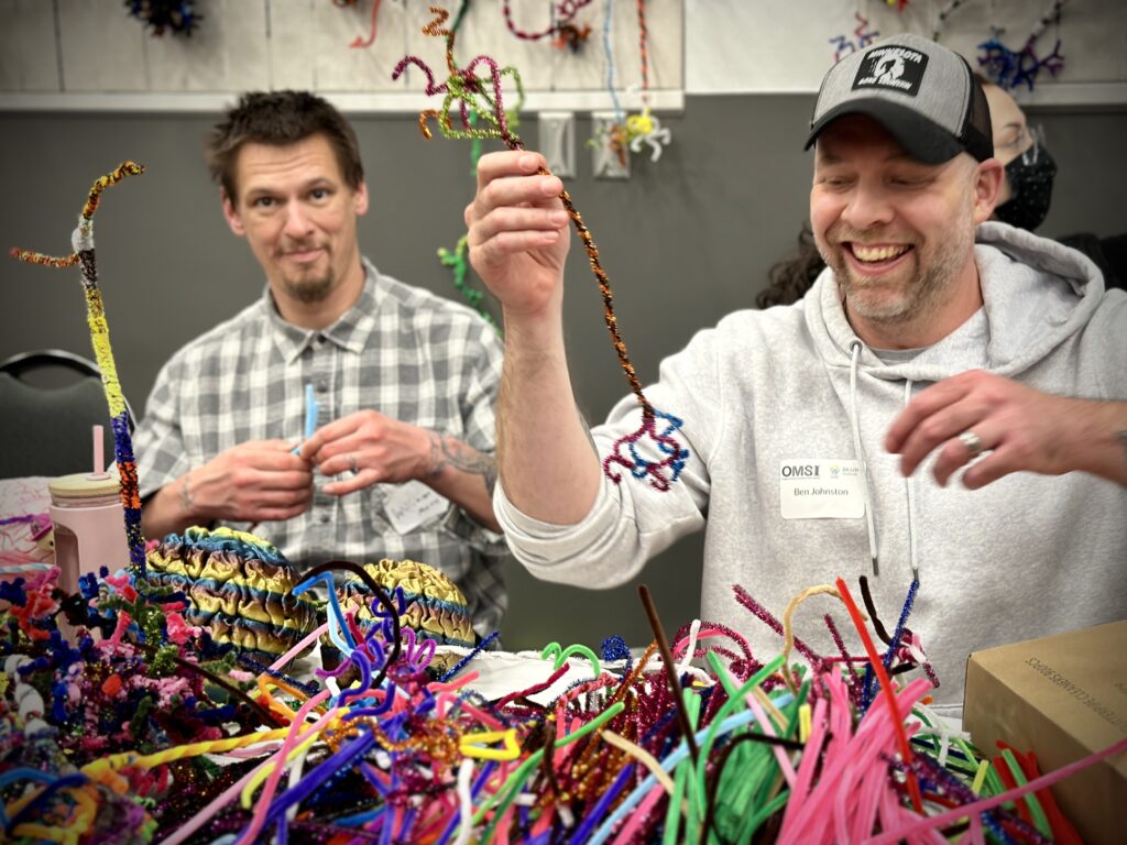
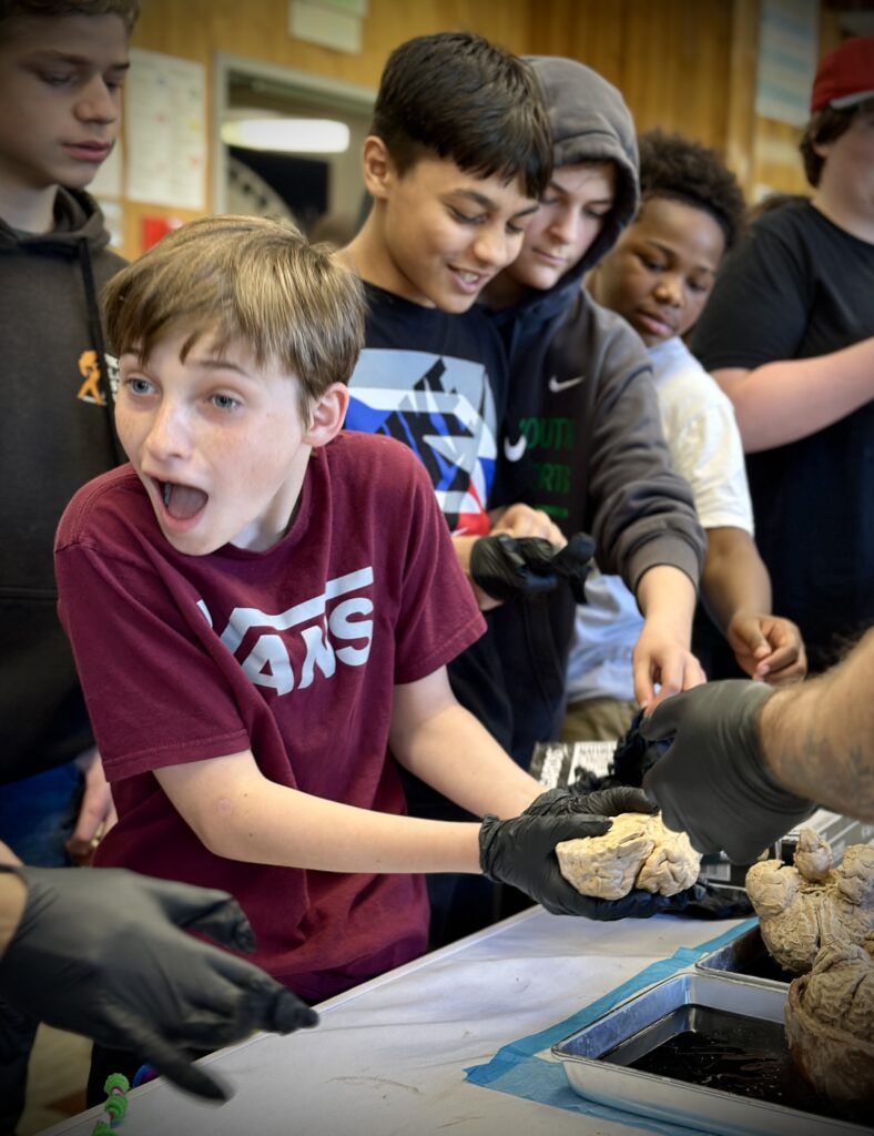
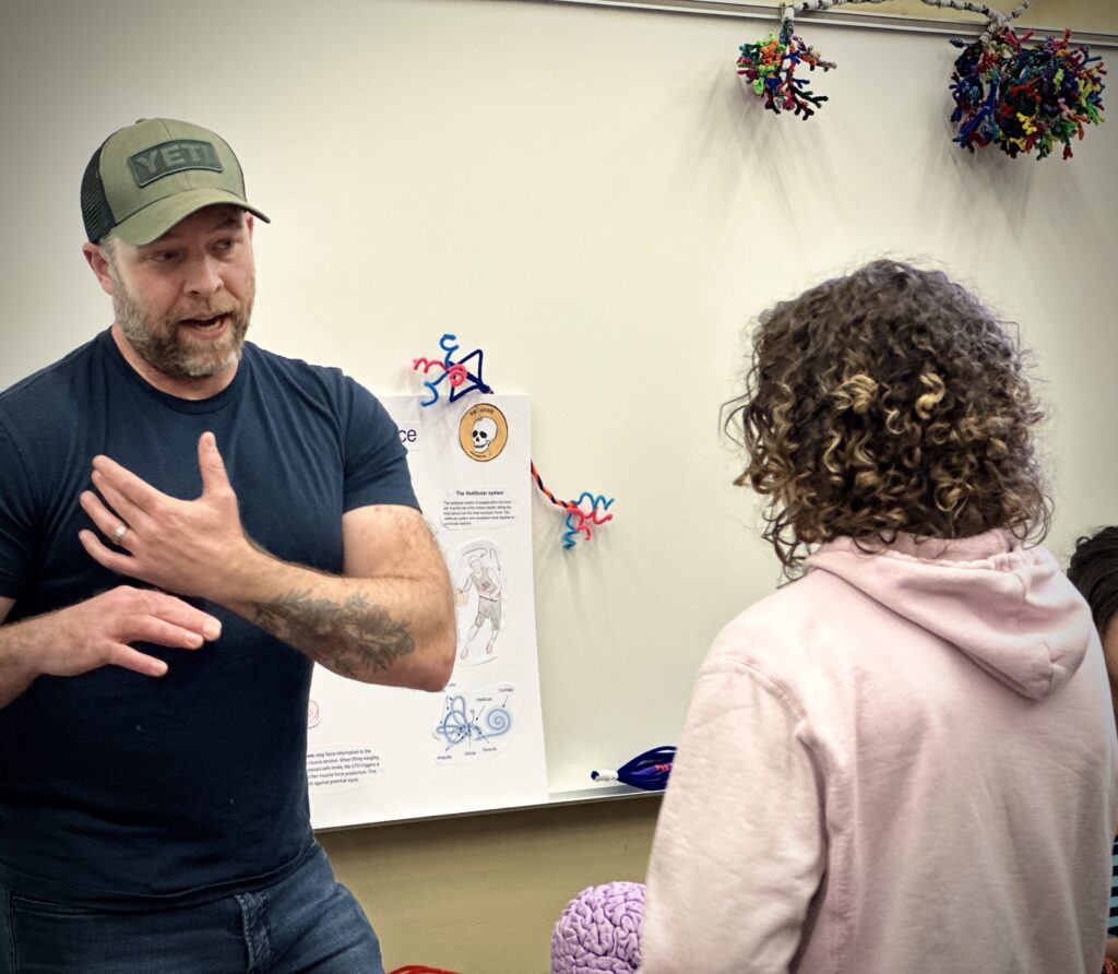
Recently, I’ve been able to join fellow students to visit OMSI (for the Brain Fair), Hosford Middle School, and Sunnyside Elementary School. During our days at these locations, we’ve been able to connect with kids on various neuroscience topics, and answer lots of curious questions – how many brain cells do we have? What does my brain do while I am sleeping? What happens if I lose a chunk of my brain?
While these kids are flexing their brains and extending their curiosity, they are also flexing their muscles. This is where neuroscience crosses over into exercise science.
Exercise science is the study of movement and the physiological adaptations and responses it produces. I pursued this area of study so I could help others improve their quality of life. The field of exercise science includes coursework in biology, kinesiology, exercise assessment and prescription, and nutrition. Exercise physiologists work in sports medicine, professional sports, gyms, and schools.
LEARN MORE: Exercise sustains the hallmarks of health
LEARN MORE: Exercise Physiology From 1980 to 2020: Application of the Natural Sciences
LEARN MORE: Research In the Exercise Sciences; Where We Are and Where Do We Go From Here
So, what do neuroscience and exercise science have to do with one another?
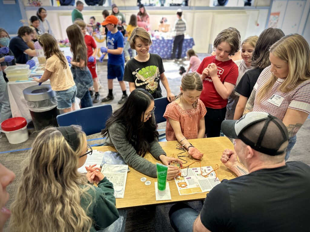
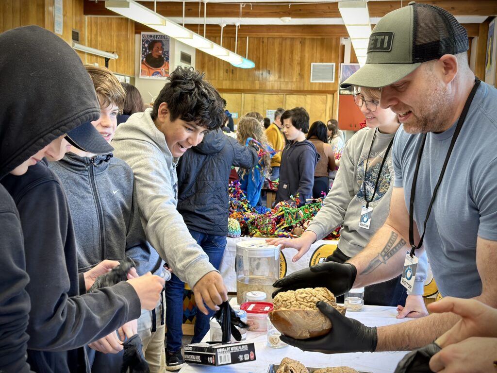
The nervous system is responsible for the processes that underlie the preparation, execution, and control of movement. During physical activity, there is a continuous flow of sensory information that is received from receptors all over the body. While observing with their eyes, using their arms and hands to pick up brains (!), and walking with their legs and feet from station to station, these kids are contracting and lengthening muscles and constantly adjusting their equilibrium.
First let’s talk about movement and muscles.
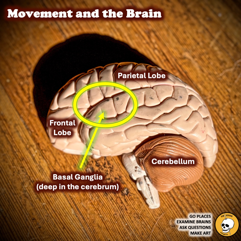
The brain has several areas associated with movement.
The frontal lobe allows us to make voluntary muscle movements. The parietal lobe is associated with spatial awareness and receives sensory information from the skin, joints, and muscles. The cerebellum is important for the coordination of movement as well as for maintaining balance. The basal ganglia (deep in the cerebrum) help determine when we start and stop movements, and how fast we go.
LEARN MORE: New insights into the brain’s motor cortex
LEARN MORE: Movement: How the Brain Communicates with the World
LEARN MORE: Neural Centers Responsible for Movement
LEARN MORE: Learning through mistakes!
Proprioception is the continuous flow of sensory information from receptors located in the muscles, tendons, joints and inner ear regarding movement and body position.
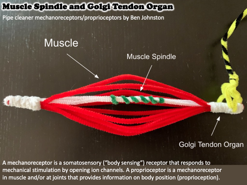
Our skeletal muscles also contain several types of specialized sensory (or afferent) receptors that send a constant flow of information to the central nervous system (CNS). Also known as mechanoreceptors, because they signal in response to mechanical movements, these specialized cells are constantly providing information to the CNS about changes in muscle pressure and length.
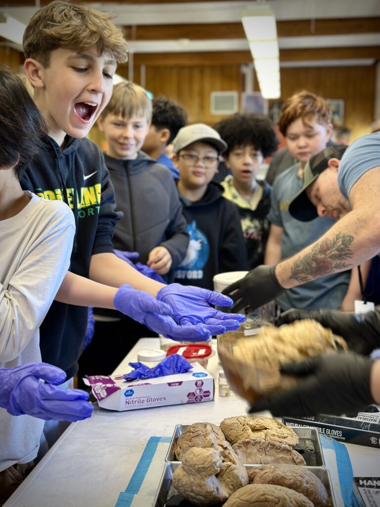
When we are active, like when we’re picking up brains with our hands, our muscles are constantly contracting and stretching. Mechanoreceptors known as muscle spindles provide sensory information about the relative length of a muscle, while cells called Golgi tendon organs monitor muscle tension. The special properties of these receptors allow them to detect and regulate muscle stiffness and protect our muscles from damage.
LEARN MORE: Proprioception and the predictive sensing of active self-motion
Muscle spindles and Golgi tendon organs!
Muscle spindles are found in large numbers in most human muscles. The muscle spindle functions as a muscle length detector. If the muscle is stretching too far, muscle spindles will trigger your muscle to contract to prevent injury to the muscle. This reflexive contraction is called the stretch reflex.
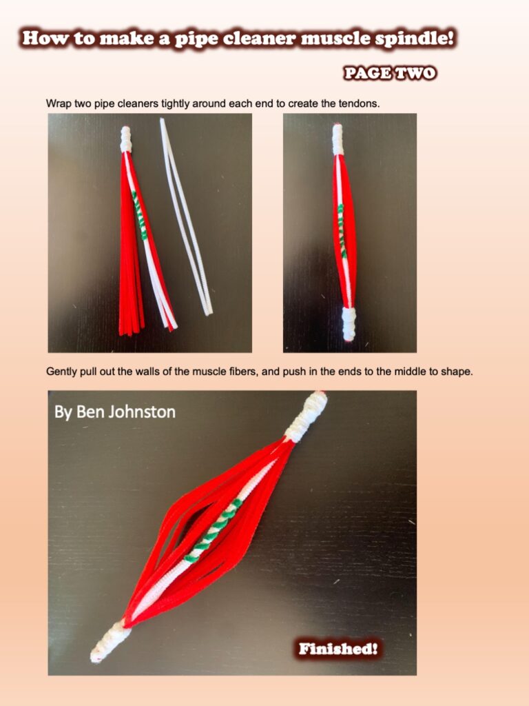
Make your own muscle spindle!

LEARN MORE: Muscle spindles and their role in maintaining robust locomotion
LEARN MORE: Muscle spindles and their role in maintaining robust locomotion
LEARN MORE: Pipe Cleaner Brain Cells!
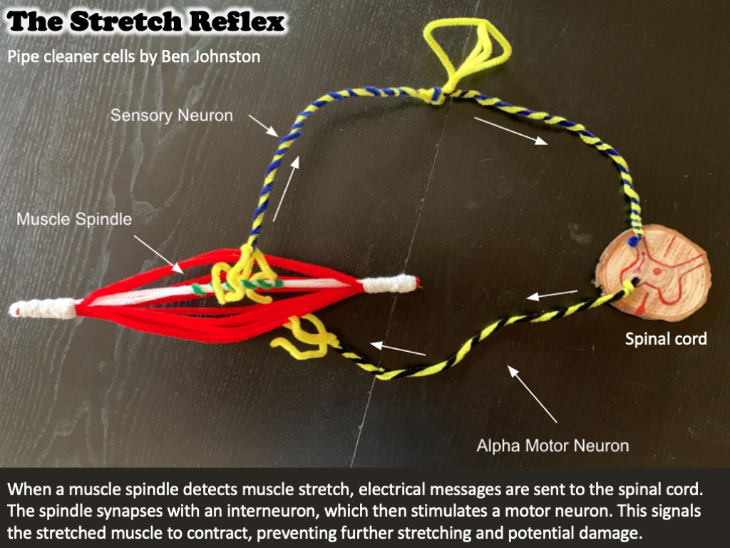
Muscle spindles are important for the stretch reflex
When it detects the stretch of a muscle, a muscle spindle sends action potentials to the spinal cord. The sensory neuron synapses with an interneuron, which then stimulates a motor neuron. The motor neuron signals the stretched muscle to contract, preventing further stretching and potential damage.
LEARN MORE: The Spinal Cord Circuitry Underlying Muscle Stretch Reflexes
LEARN MORE: The neurophysiology of tone: the role of the muscle spindle and the stretch reflex
What do the Golgi tendon organs do?
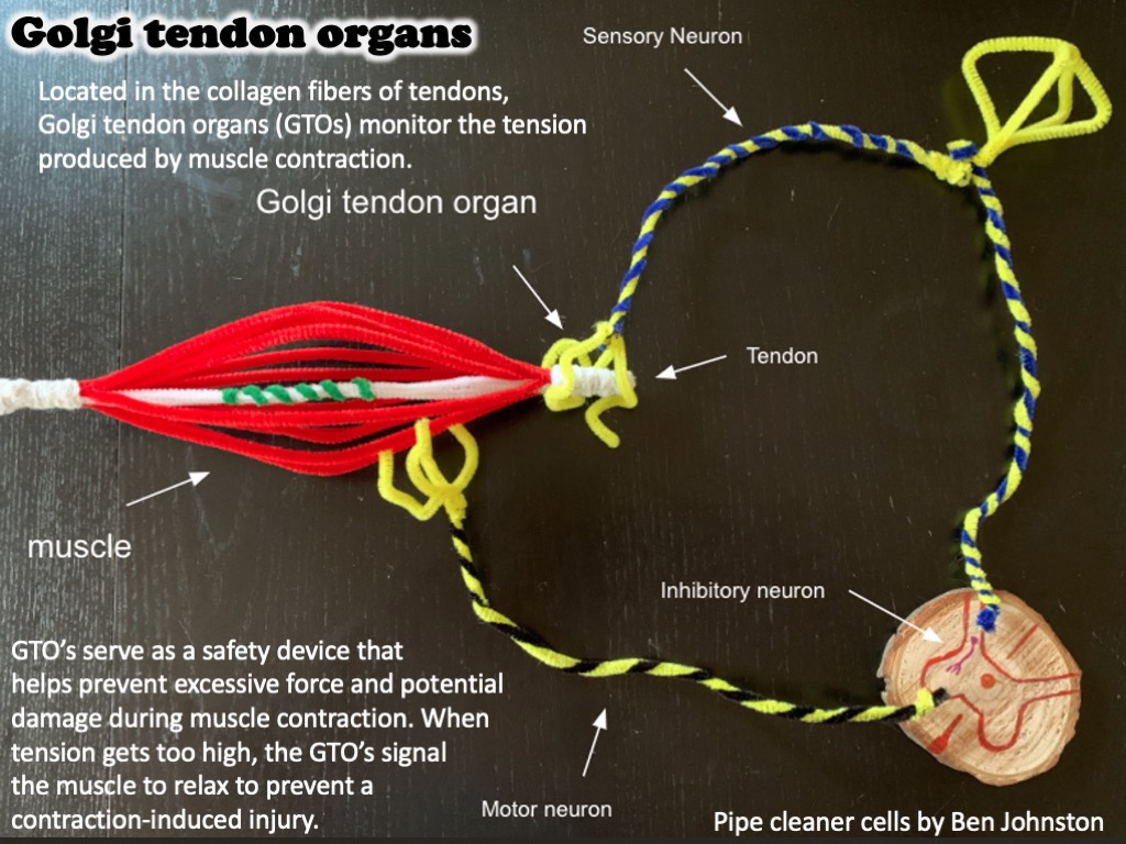
Golgi tendon organs (or GTOs) help prevent excessive force and potential damage during muscle contraction. Located in the collagen fibers of tendons, GTOs continuously monitor the tension produced by muscle contraction. When the tension gets too high, the GTOs act as a safety device, signalling the muscle to relax to prevent a contraction-induced injury in a process known as autogenic inhibition.
LEARN MORE: Molecular correlates of muscle spindle and Golgi tendon organ afferents
LEARN MORE: Distributed force feedback in the spinal cord and the regulation of limb mechanics
LEARN MORE: Functional properties of the Golgi tendon organs
LEARN MORE: The Golgi tendon organ: a review and update
Now, let’s talk about balance and equilibrium
Another vital aspect to movement is balance and spatial orientation. When the students look from the brain specimens to other students then to teacher and back again, they are making skilled positional movements based partly on sensory input from their vestibular apparatus.
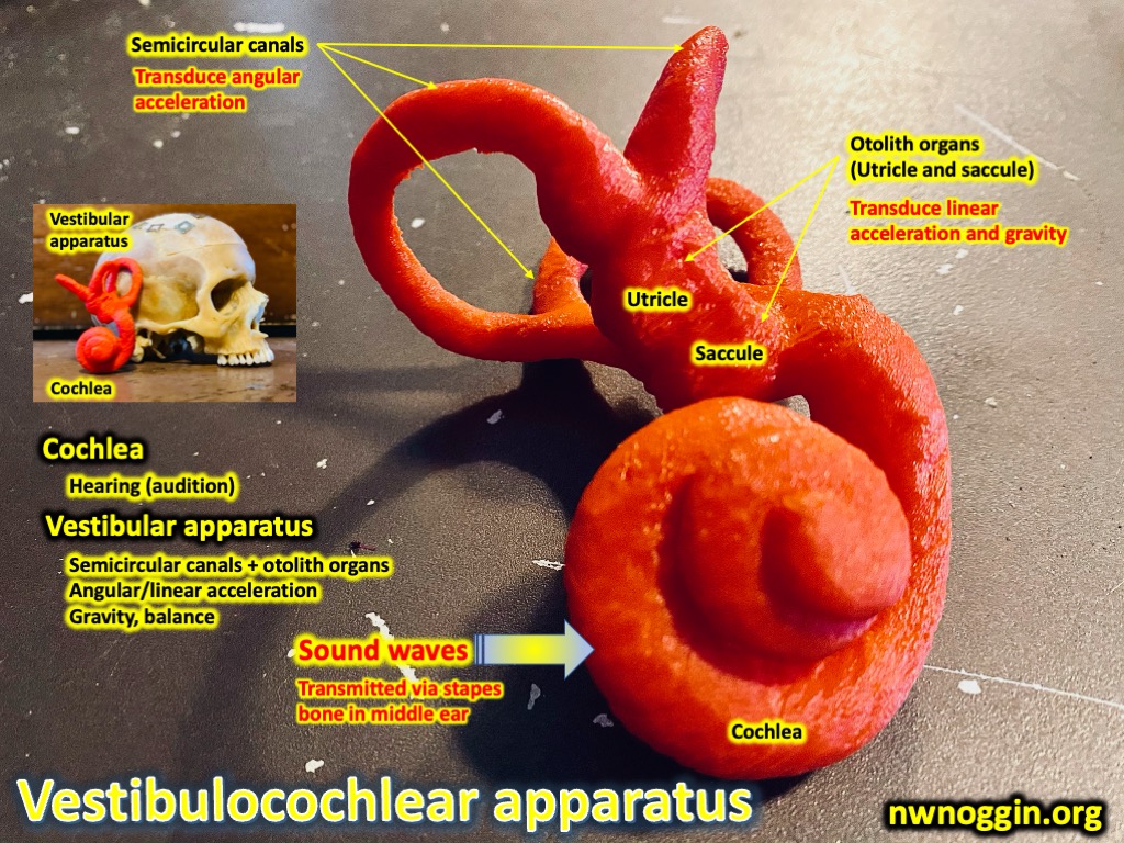
The vestibular apparatus is an organ located in the inner ear, connected closely with the cochlea, a structure that is essential for hearing. The specialized receptors within the organ are sensitive to change in head position and head movement. During physical activity, the vestibular apparatus informs head and eye movement to maintain balance and to visually track the consequences of movement.
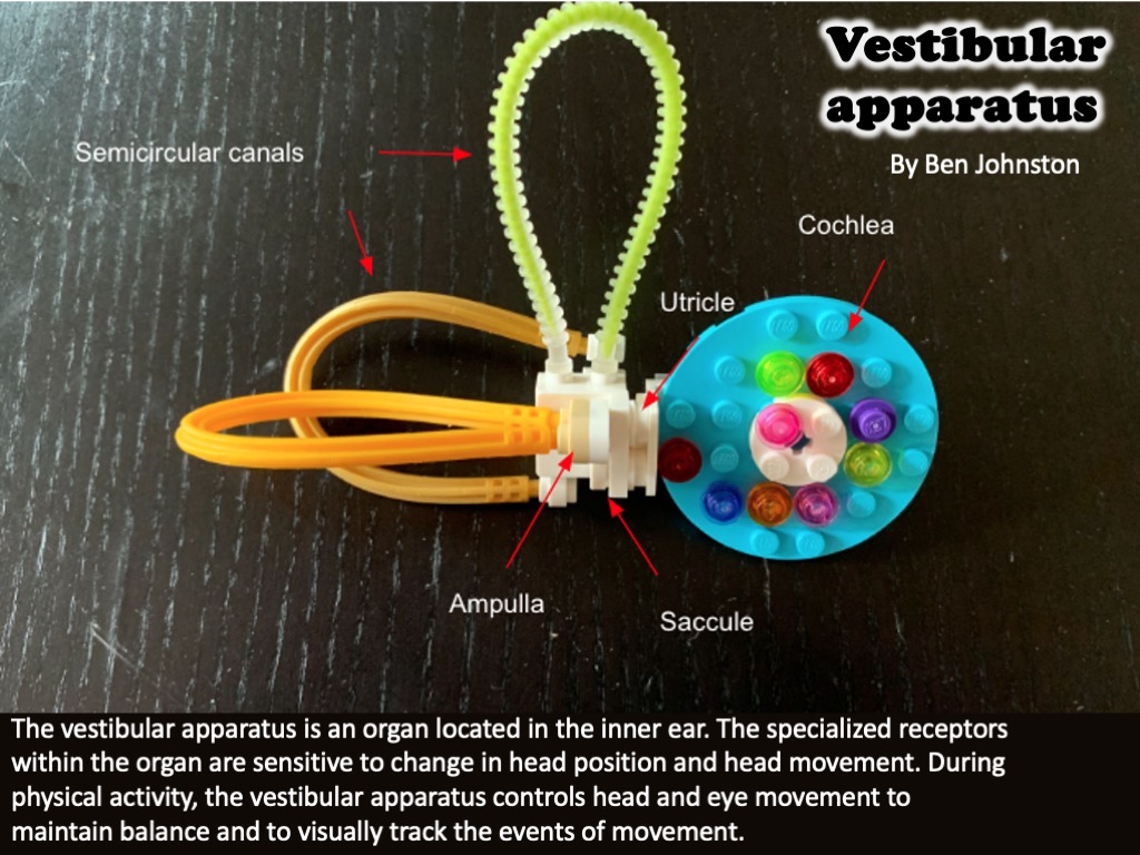
The otolith organs (the utricle and the saccule) and the three semicircular canals of the vestibular apparatus play a key role in the process of balance. The semicircular canals are filled with endolymph fluid and lie at right angles to one another. This allows each canal to be stimulated by a different directional movement of the head.
At the end of each canal is a small bulb-like structure (the ampulla) that contains the sensory afferents known as “hair cells.” The hair cells are topped by a gelatinous cone-shaped cap called the cupula. The lightweight cupula floats in the endolymph that fills the semicircular canals.

LEARN MORE: Anatomy of the vestibular system: a review
When the head rotates, hair cells within the ampulla are moved, and stimulate nearby nerve receptors to send signals to the brain. This triggers the responses necessary for maintaining balance when the head or body is suddenly accelerated in a particular direction.
There are also two additional vestibular organs called the utricle and the saccule. A patch of hair cells lies inside both of these chambers. The tips of their hair cells are covered by a gelatin-like material. Within the gelatin material are heavy mineral (limestone!) crystals called otoliths.
The otoliths remain level on the hair cells when we’re not moving, creating minimal stimulation. When the head tilts forward or when the entire body accelerates forward or backward, the membrane and the otoliths shift, stimulating the hair cells. This, in turn, stimulates nearby receptors of the vestibular nerve to send impulses to the brain, producing a sense of our changing position.
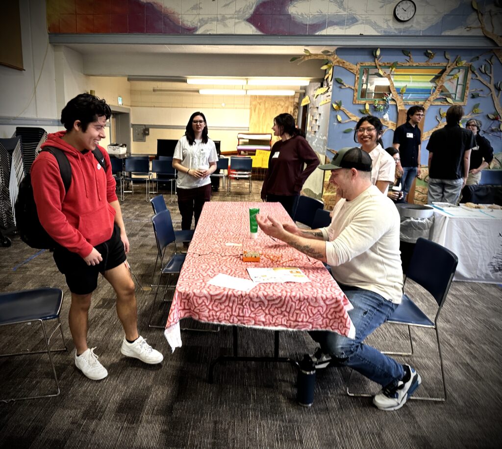
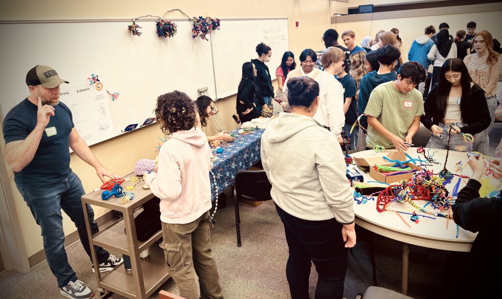
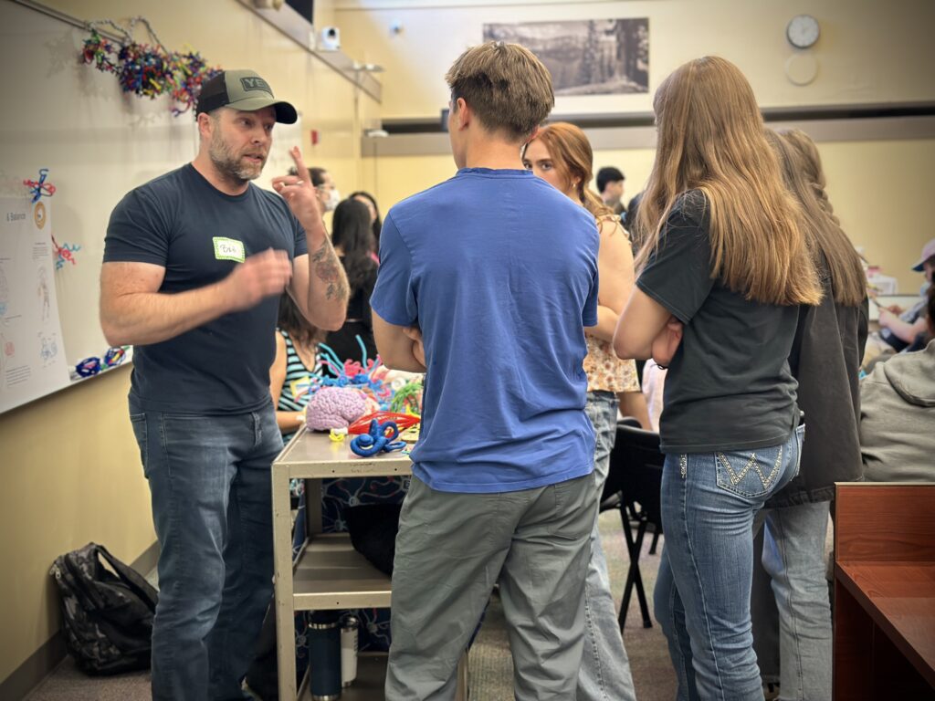
Outreach at David Douglas High School
At David Douglas High School, I had the opportunity to bridge the disciplines of neuroscience and exercise science. I talked with students about the areas of the brain associated with movement, and we also explored the special sensory receptors that are involved in sending important information to the central nervous system about muscle tension, stretch and balance.

While talking with high school students, I was asked some great questions!
How does exercise benefit the brain?
There are many ways that regular exercise can benefit our brain health.
Regular exercise helps us sleep better at night. While we are sleeping, cerebrospinal fluid flushes toxic, memory-impairing proteins from the brain! In addition, regular sleep improves our mood, helps us think clearly, and improves learning.
Regular exercise also increases mood regulating neurotransmitters resulting in decreased stress. Mental health also improves in individuals who exercise more regularly, especially when social engagement is added. Regular long walks with a good friend is fantastic for mental health.
LEARN MORE: Health Benefits of Exercise
LEARN MORE: Real-Life Benefits of Exercise and Physical Activity
LEARN MORE: The Effect of Physical Activity on Sleep Quality and Sleep Disorder
LEARN MORE: How Sleep Clears the Brain
What is the worst kind of sports injury?
My reply to this was “any injury that results in brain damage is going to be pretty bad.”
Concussions are mild traumatic brain injuries that affect brain function and are very common in certain types of contact sports.
LEARN MORE: Concussions and their consequences: current diagnosis, management and prevention
LEARN MORE: A Bang to the Brain
Can you go blind if you hit your head too hard?
Post traumatic visual loss is common after head trauma, including sport related injuries. However, this is even more common following direct injuries to the eyes.
LEARN MORE: Post-traumatic visual loss
LEARN MORE: An insight into the vision impairment following Traumatic Brain Injury
During my time with Northwest Noggin I’ve learned so much from fellow volunteers and from the young people I met in schools. Sharing my knowledge, learning from questions, making brain art, and connecting with my community was such a great experience.