Post (and illustrations) by Ben Wall, a senior at Portland State University graduating in June, 2024 with a Bachelor of Science in Psychology and a minor in Interdisciplinary Neuroscience. Originally from Hood River, Oregon, Ben spent ten years in forestry and two years as a case worker for the homeless community prior to returning to school in 2021.

The Push for New Mental Health Tools
In the spring of 2021, I was living at my parents’ house and awakening from a long recovery from a manic episode and subsequent depression. Mania is a state of extreme mood elevation that teeters on delusions and detachment from reality. It was the second manic episode of my life, but the first one observed by doctors, which resulted in a diagnosis of bipolar disorder.

LEARN MORE: Mania
LEARN MORE: What is bipolar disorder?
Bipolar disorder is characterized by periods of mania and depression; high and low extremes of mood. After six months of one of the worst depressions of my life and wrestling with the diagnosis, I decided to try medication and therapy…again. My attempts with various medications over the prior two years had been frustrating and fraught at times, but I now had a more informed diagnosis. Medication brought me back to life, and therapy helped me come to terms with my diagnosis and recover my spirit. Feeling semi-normal again was an incredible relief, but it wasn’t good enough.
LEARN MORE: Diagnosis and Treatment of Bipolar Illness in the Primary Care Office
LEARN MORE: Pharmacological Treatment of Bipolar Depression: Current and Emerging Options
I had cycled through depressions since I was a child, and over ten years of my adult life had been raggedly defined by highs and lows that led to many new starts. An earlier diagnosis and effective treatment would have saved me a lot of grief. Many bipolar diagnoses don’t occur until after an incident that involves police and/or doctors, and at that point, a life has likely already been derailed. People who suffer from mental illness are often misdiagnosed and go through years of trying different medications and therapies before an effective treatment provides stability to their lives, and some never find relief.
As I climbed out of my last depression, I had endless questions about my mind and brain, and a strong desire for better mental healthcare diagnostics and treatment options. I had little idea of my direction, but I knew that the starting point was education, and I enrolled at Portland State University that fall to pursue a degree in psychology.
Three years later, I find myself working in labs at Oregon Health & Sciences University pursuing objectives that were previously only tangible in my dreams: finding new and more effective ways to diagnose and treat mental illnesses.

Studying human behavior to prevent suicide

On Mondays and Wednesdays I spend time in the Huber Lab in OHSU’s Center for Mental Health Innovation. Led by Dr. Rebekah Huber, the lab is studying youths with bipolar disorder who have recently attempted suicide. The rate of suicide attempts by adolescents with bipolar disorder is 50 times higher than that of the general adolescent population, and there is an urgent need for effective intervention strategies. The objective of this study is to identify early warning signs in adolescents with bipolar disorder that indicate they are at risk of attempting suicide.

LEARN MORE: Prevention of suicidal behavior in bipolar disorder
Study participants are monitored for their mood, sleep, mental task performance, and physical activity patterns, as well as any occurrences of suicidal thoughts for 30 days. Participants begin the study with a three hour interview to determine their eligibility and ensure that they understand what their voluntary participation entails. Participants are asked about relationships with family and friends, school and extracurricular lives, experience of mental illness, and history of suicidality.
Week one of the study begins with an assessment of the participant’s cognitive abilities using a set of mental task tests. One cognitive ability of particular interest is cognitive control, which refers to the ability to perform a task despite distracting impulses, such as being able to read an article despite hearing text message notifications. An example of one of the tests used is the Stroop Task, which involves reading a list of color words that are printed in a different color while being timed.

LEARN MORE: The Stroop Color and Word Test
The assessment gives a standard measure of the participant’s cognitive control, and during the study they will receive daily mental task tests via a mobile phone app that will track their cognitive functioning, as well as other cognitive abilities. The app also administers random daily check-ins that ask about the participant’s mood and thoughts of suicide. The study will use this data to explore possible correlations between mood fluctuations, cognitive control, sleep, physical activity, and suicide risk among the participants.
Most of my work in the lab has been to read research articles about bipolar disorder and compile a literary review, or a summary of the research. The topic of my review is abnormalities in brain function in bipolar disorder and treatments that potentially target those dysfunctions. Brain imaging studies have identified various differences in functioning between healthy human brains and the brains of individuals with bipolar disorder. Theories abound about how abnormalities in brain function might affect an individual with bipolar disorder, and what they might mean for treatment and diagnostic options.
LEARN MORE: Functional disconnection between subsystems of the default mode network in bipolar disorder
LEARN MORE: Epidemiology of suicide in bipolar disorders
The search for a genetic basis of schizophrenia
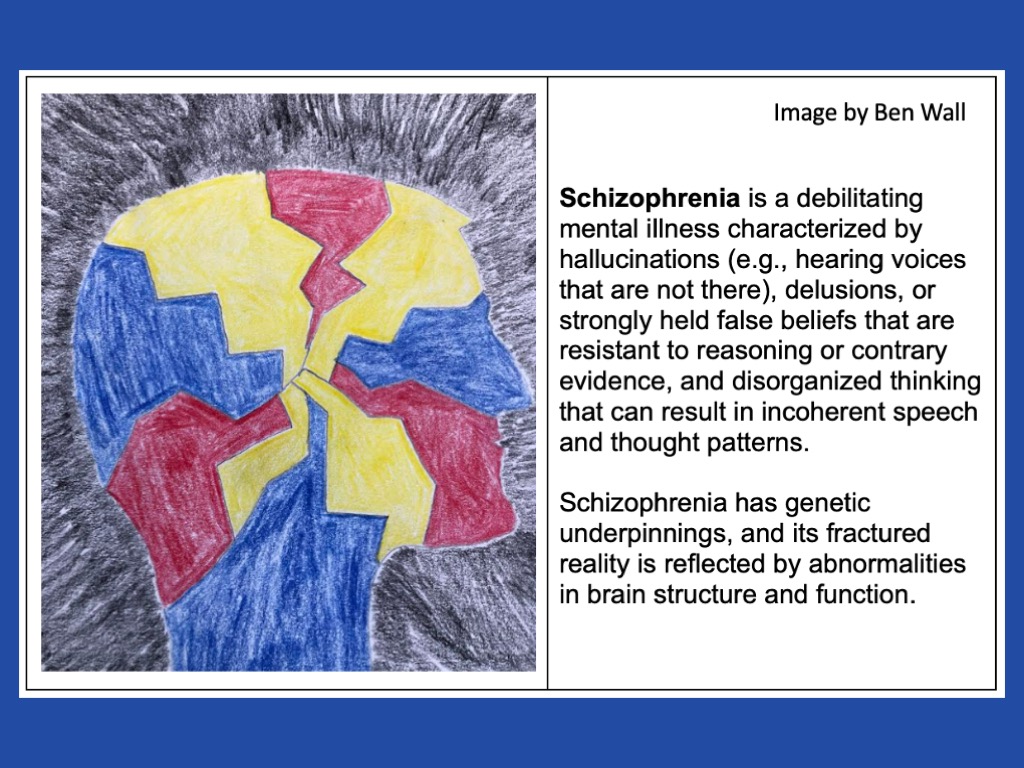
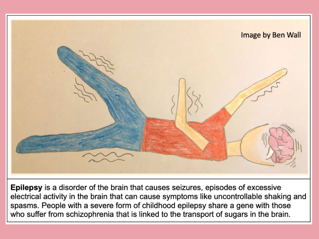
Tuesdays and Fridays I spend time in the lab of Dr. Robert Mealer at OHSU. Dr. Mealer studies genes that increase the risk of developing schizophrenia, as well as childhood epilepsy.

The goal is to identify a biological mechanism for how these genes contribute to brain disorders. To answer these questions, he studies the brains of mice that have been genetically engineered to have this gene. These mutant mice are ordered custom-made from a laboratory, along with mice of the “wild type,” or the genetically unaltered versions.

LEARN MORE: Schizophrenia: Overview and Treatment Options
LEARN MORE: Neurobiology of Schizophrenia: A Comprehensive Review
LEARN MORE: Genetics of schizophrenia (Review)
LEARN MORE: What are epilepsy and seizures?
LEARN MORE: The Genetics of Epilepsy
The Mealer Lab has many different mouse specimens to experiment with, and the first step is to sort out the mutant mice and the wild type mice. To distinguish the genetic differences of the mice, a polymerase chain reaction, or PCR test, is performed. PCR is a common and versatile lab technique that magnifies sequences of DNA so that they can be studied. The multi-step process begins with soaking a tissue sample, like the tip of a mouse tail, in a chemical solution and heating it to near boiling for two minutes. This step extracts DNA from the tissue. Next, the DNA needs to be multiplied exponentially so that it can be studied. The extracted DNA has a mixture added to it that will locate and duplicate the specific segment of DNA that distinguishes the mutant mice from the wild type. The concoction, all mixed in a tiny plastic tube half the size of a pinkie finger, is then placed in a machine that takes it through dozens of heating and cooling cycles. With each cycle, the two strands of molecules that form DNA separate and then duplicate. The multiplied DNA is then mixed with a dye that will make it visible with UV light when photographed with a specialized camera.

LEARN MORE: What is PCR?
LEARN MORE: Research Techniques Made Simple: Polymerase Chain Reaction (PCR)
The final step is to measure the DNA using a technique called gel electrophoresis.
A firm block of gel the size of a small paperback book is submerged in liquid in an electrically charged box. The DNA samples are injected into the gel in a row like the starting line of a race, and when the electrical current is turned on, the charge pushes the DNA molecules through the gel. The current is left on for 40 minutes, and in that time, DNA molecules travel different distances through the gel. The smaller the molecule, the farther it pushes through the gel. The mutants and wild types are then identified based on how far their DNA samples traveled through the gel.

LEARN MORE: Agarose Gel Electrophoresis for the Separation of DNA Fragments
LEARN MORE: When a Western Blot Goes SOUTH
LEARN MORE: Southern blotting
As a novice, running a PCR test takes me four to five hours and several pages of notes. I quickly became acquainted with pipettes, which are like syringes that precisely measure miniscule volumes of liquid, and learned the different sizes and functions of various test tubes. It took me three tries on my own before I performed a successful PCR test. One error at any point in the process can result in a faulty experiment, but it likely won’t be known until the experiment is complete. Then begins the process of combing through notes taken during the experiment and hunting for what might have gone awry.
Once it has been determined whether the mice are mutants or wild types, their brains are ready to be probed in search of differences caused by the mutations. To begin the probe we would view brain slices of the mice under a microscope. This was no ordinary microscope, though, and it would be an hours-long process of preparing the samples for viewing.

Of interest is the transport of various types of sugar within the brain.
It is known that the mutant mice transport sugars differently than the wild type, and a microscope can be used to view where the sugars are located in the brain. To do this, a razor-thin cross-section of the brain is soaked in a liquid containing lectins. Lectins are plant proteins that bind to very specific sugars, so the lectins will attach to the sugars in the brain slice. The lectins contain a fluorescent marker that is visible when viewed through a microscope that shines specific wavelengths of light through the sample.

Dr. Mealer describes his investigation as a 20 to 30 year project.
The examination of biological abnormalities in the brains of individuals with schizophrenia, beginning with the examination of mice with similar abnormalities, will hopefully one day lead to new methods for diagnosing schizophrenia, and possibly new treatments.
LEARN MORE: Glycobiology and schizophrenia: A biological hypothesis emerging from genomic research
LEARN MORE: The schizophrenia-associated variant in SLC39A8 alters protein glycosylation in the mouse brain
LEARN MORE: The schizophrenia risk locus in SLC39A8 alters brain metal transport and plasma glycosylation
LEARN MORE: N-glycome analysis detects dysglycosylation missed by conventional methods in SLC39A8 deficiency
Mining big data for answers to mental illness
My Thursdays are spent in OHSU’s Intergenerational Neuroimaging Lab, led by Dr. Alexander Dufford. Dr. Dufford uses brain imaging, particularly magnetic resonance imaging (MRI), to study mental illnesses.
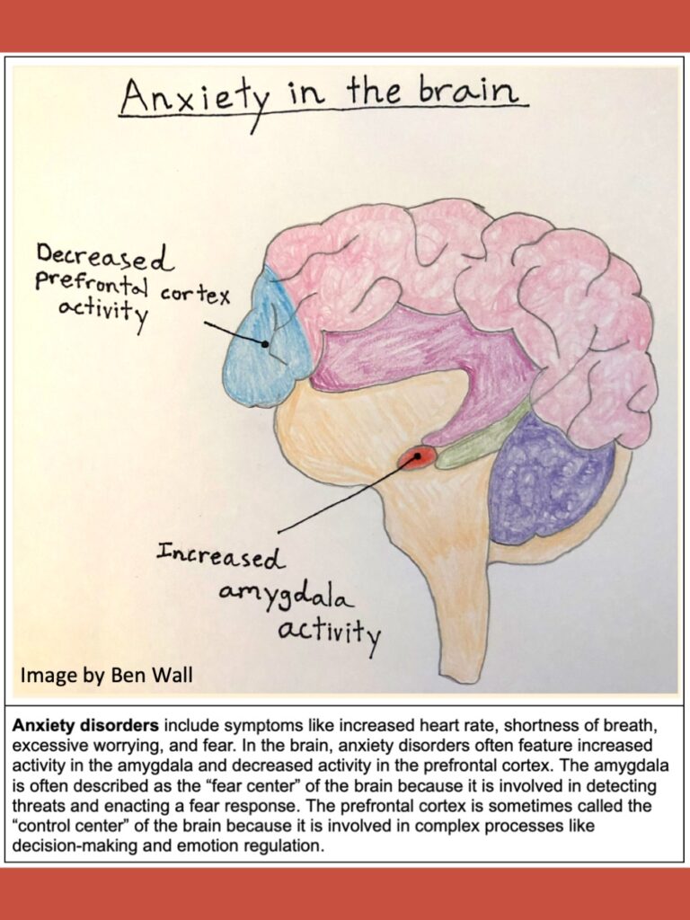
The current project is examining connections within the brains of youth between the ages of 5-21 who have anxiety disorders, and then exploring how the brain connections in that group compare to the brains of a group of preschoolers with anxiety disorders. The objective is to find out if anxiety disorders in youth can be predicted by examining their brain connections at preschool age, which could enable earlier diagnosis.

LEARN MORE: What is anxiety?
LEARN MORE: Anxiety disorders
The Intergenerational Neuroimaging Lab has taken me down a learning path that I did not foresee: learning computer software programming, or coding. Many researchers use a computer programming language called Python to record and analyze data. It can be used to create spreadsheets like Microsoft Excel or Google Sheets, but it can manage enormous amounts of data in comparison. For example, say a data table includes brain images for 4,000 people, and included in the table are each person’s age, sex, race, and mental health diagnosis. Perhaps the study only wants to examine the brains of hispanic males between the ages of 35-40 who have been diagnosed with major depressive disorder. Using Python, a code can be written that will sort through the thousands of lines of data and retrieve only the data of interest. The amount of data that can be sorted and analyzed by Python in a few minutes could take hours on a common program like Microsoft Excel, assuming it doesn’t crash the program in the process.

LEARN MORE: Python in neuroscience
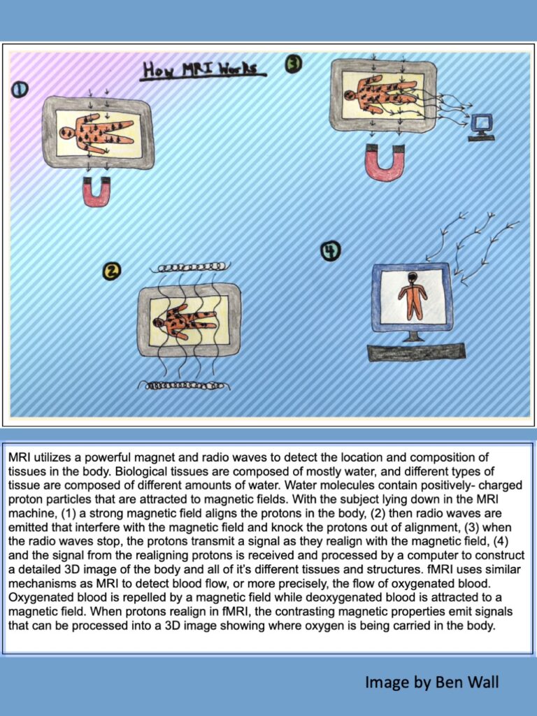
MRI produces images of the brain and its structures.
MRI can differentiate between different brain tissue types to produce a detailed digital 3D model.
Then there is functional MRI, or fMRI, which senses blood flow and indicates where cellular activity is occurring in the brain.

fMRI images can be taken to show blood flow when a person is merely laying in the imaging machine and their brain is “at rest,” or blood flow can be imaged when the person is performing a task, such as the Stroop Task that the Huber Lab uses. Neuroscience researchers around the world collaborate by sharing their imaging data on large databases like OpenNeuro which is accessible to anyone.

Using open access databases like OpenNeuro and the analytical tools of Python, the Intergenerational Neuroimaging Lab can study thousands of brains. Brain structure and function associated with different mental illnesses can be examined using MRI and fMRI, and findings can be compared across generations and across different age groups to learn about the development of mental illnesses. By learning about the development of mental illnesses, scientists can find new techniques for earlier and more accurate diagnosis.
LEARN MORE: The Intergenerational Neuroimaging Lab
LEARN MORE: Scientific Research and Big Data
Forging a Path Ahead
Science is a collaborative effort.
A scientist might have an idea, a scientific hypothesis, another scientist has tools that can help investigate the idea, and then a third scientist maybe has an idea for investigating the hypothesis from a different angle. Science is always changing, progressing, and correcting past mistakes.
The work of Dr. Huber, Dr. Mealer, and Dr. Dufford is part of a collaborative effort to improve the quality of life for those living with mental illness.
The Huber Lab is investigating early warning signs of suicide in youths with bipolar disorder so that lives can be saved. The Mealer Lab is investigating the genetic underpinnings of schizophrenia so that people can be diagnosed sooner and when the disease is more treatable. The Intergenerational Neuroimaging Lab utilizes MRI and fMRI along with big data analytics to study brain connections in youth with anxiety disorders. By comparing brain structures and functions across different age groups, they hope to identify early indicators of anxiety disorders that enable earlier and more accurate diagnosis.
These interdisciplinary efforts at OHSU are part of a global collaboration of neuroscientists who are tirelessly striving for advancements in the diagnosis and treatment of mental illnesses, and ultimately to improve the quality of life for those living with mental illness.


