Post by Lorenzo Nungaray, undergraduate in Biology pursuing an Interdisciplinary Neuroscience minor at Portland State University. Lorenzo works as a research technician in the Sleep & Health Applied Research Program (SHARP) at OHSU, under the direction of Dr. Miranda Lim.

LEARN MORE: Lorenzo Nungaray, SHARP Lab @ OHSU
“Each night, when I go to sleep, I die. And the next morning, when I wake up, I am reborn.”
— Mahatma Gandhi
Sleep is a fundamental aspect of human life, yet its importance has often been overlooked and underestimated. Most people have experienced the grogginess of a sleepless night or the refreshing feeling of a good night’s rest, but the significance of sleep extends far beyond these subjective experiences.
Is there a reason that we spend so much time sleeping and if so, what is that reason?
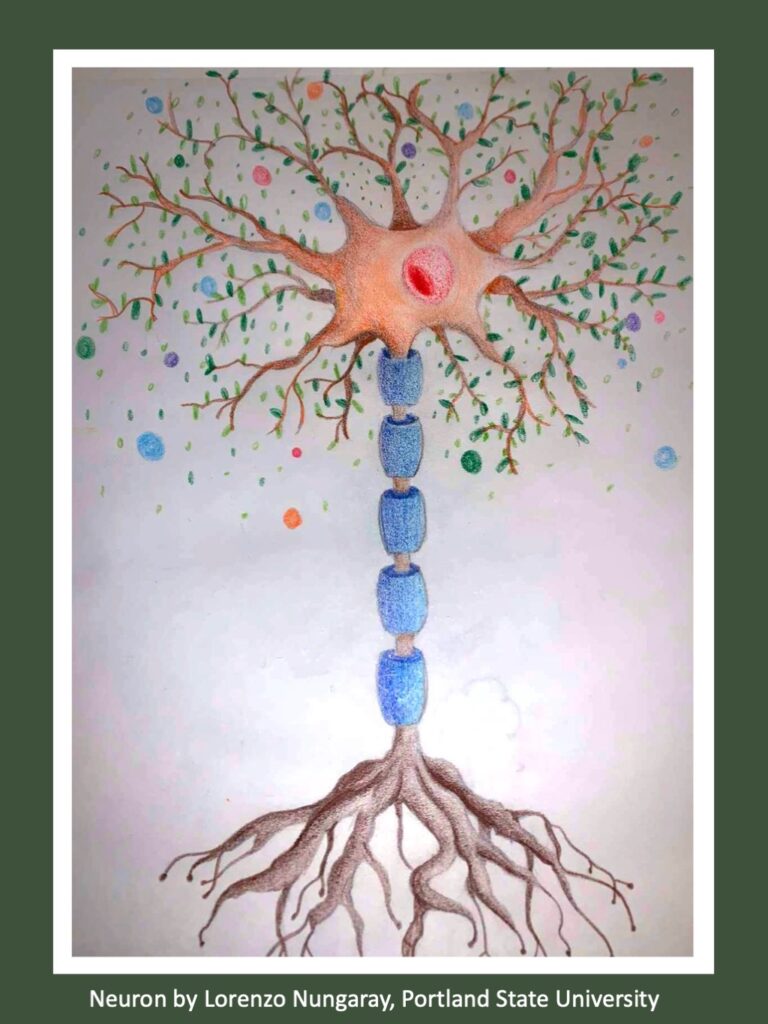
Recently, I have had the privilege to work at SHARP (Sleep and Health Applied Research Program) with some amazing people who want to answer these same questions. My time while working for SHARP has led me to find some amazing facts about sleep and why it plays such a vital role in our lives.
SHARP logo
Sleep is a complex and essential process that is divided into distinct stages.
These sleep stages are categorized into two main types: Rapid Eye Movement (REM) sleep and Non-Rapid Eye Movement (NREM) sleep. The sleep cycle typically consists of multiple iterations of these stages throughout the night, with each stage serving specific functions.

The image above comes from sleep tracking done on myself as part of my time at the lab.
The device used is called a sleep mat which is placed under an individual’s mattress to monitor sleep cycles. This device measures the time spent asleep and divides your total sleep into each stage of sleep (Wake, Non-REM, and REM).
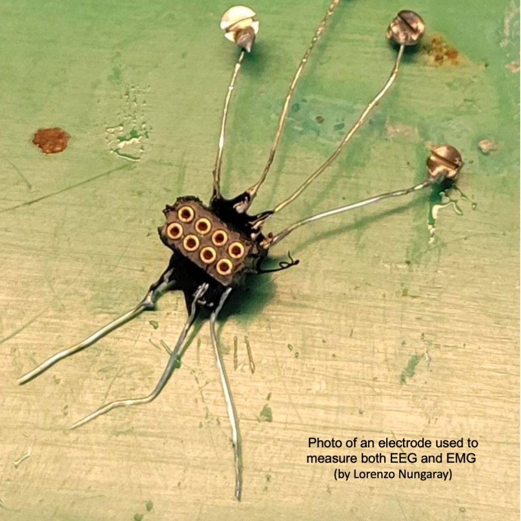
Sleep stages can be identified through the use of EEG or electroencephalogram which measures brain waves and activity and EMG or electromyogram which measures muscle activity. These values can be measured by attaching devices called electrodes to mice and recording their sleep and wake cycles.
LEARN MORE: Electroencephalography
LEARN MORE: What happens during an electroencephalogram (EEG)?
NREM (Non-REM) Sleep
NREM sleep is further divided into four stages, each with its own set of characteristics.
NREM Stage 1 is the transitional phase from wakefulness to sleep.
NREM Stage 2 is the most common sleep stage, constituting a significant portion of the sleep cycle.
NREM Stages 3 and 4 are collectively referred to as slow-wave sleep (SWS). These stages are characterized by the deepest and most restorative sleep.
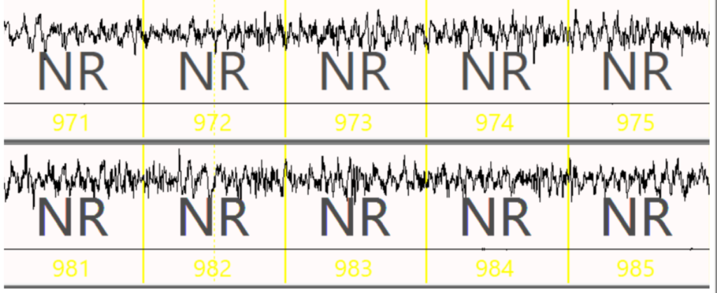
Screenshot taken from electroencephalogram (EEG) showing NREM sleep in a mouse.
LEARN MORE: Noggins in Nod: The science of sleep
REM (Rapid Eye Movement) Sleep
REM sleep is the other major type of sleep stage and is characterized by several distinctive features. REM sleep is named for the rapid and random movements of the eyes that occur during this stage. This is also the stage where most dreaming occurs.
The brain actively inhibits voluntary muscle activity leading to temporary paralysis during REM sleep. Brain activity during REM sleep is quite similar to that during wakefulness. The REM sleep stage is crucial for cognitive processes and memory consolidation.
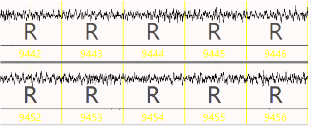
Screenshot showing REM sleep using electroencephalogram (EEG) in a mouse.
Sleep stages can be recorded and measured via EEG and EMG (the electromyogram, which measures muscle activity). The EEG (top) portion of the figure shows the electrical brain activity that determines what sleep stage the model is in while the EMG (bottom line) of the figure shows the muscle movement of the model.

Image highlighting EEG (blue) signals and EMG (red) signals during wake in a mouse model.
By comparing these two signals, we can determine if the model is asleep or awake. If they are asleep, we can determine what sleep stage they are experiencing and look for the distribution of sleep cycles in the data.
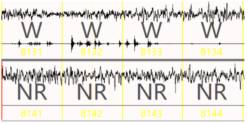
EEG/EMG showing transition from wake to non-REM sleep cycle in a mouse model.
The Glymphatic System clears out waste while you slumber
Another important function of sleep is the work done by the glymphatic system.
The glymphatic system is a recently discovered brain waste clearance system that operates during sleep. It includes a network of vessels and structures within the brain that plays a critical role in clearing waste products and maintaining brain health.
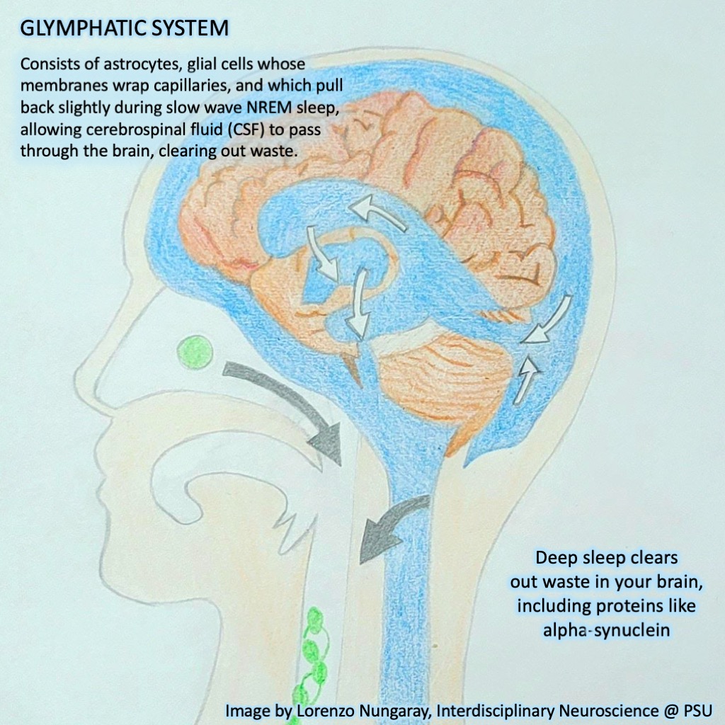
The glymphatic system primarily consists of glial cells, specifically astrocytes, which are the most abundant glial cells in the central nervous system.
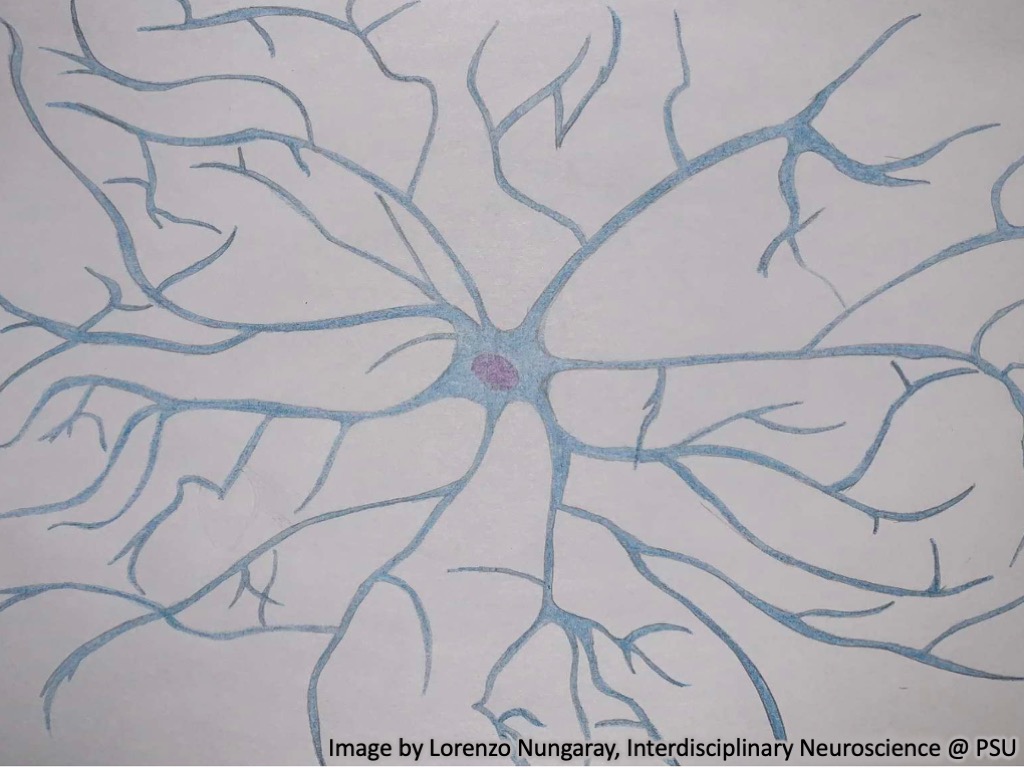
Astrocytes play a central role in maintaining the brain’s microenvironment and providing support for neurons. The glymphatic system works in concert with the perivascular space, a network of channels surrounding blood vessels in the brain. Cerebrospinal fluid (CSF) is pumped through these perivascular spaces, flushing out waste products and toxins. This CSF influx is facilitated by the expansion of interstitial spaces during sleep, allowing for efficient waste removal.
“I’ve always envied people who sleep easily. Their brains must be cleaner, the floorboards of the skull well swept, all the little monsters closed up in a steamer trunk at the foot of the bed.”
— David Benioff
What is alpha-synuclein?
The SHARP lab has taken an interest in a protein called alpha-synuclein.
Under normal conditions, alpha-synuclein would aid in neurotransmitter reuptake as well as many other functions in synaptic vesicles of neurons.
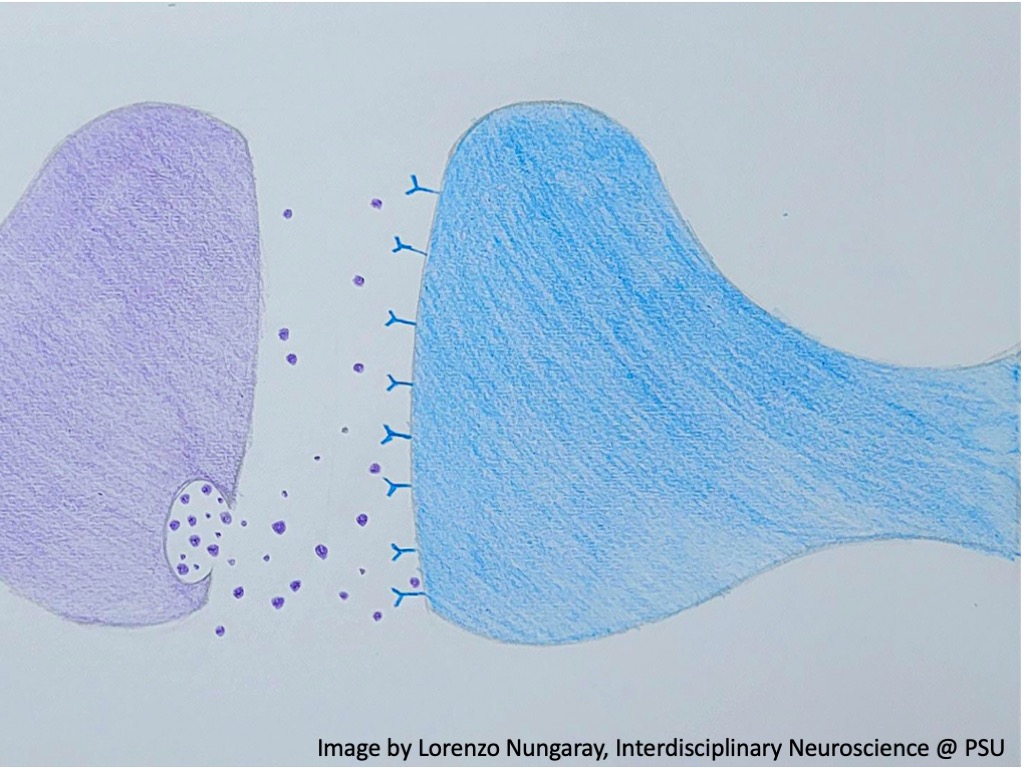
However in Parkinson’s disease, alpha-synuclein takes on a pathological form, forming insoluble aggregates known as Lewy bodies and Lewy neurites. These aggregates accumulate in various regions of the brain, including the substantia nigra, a region associated with motor control.
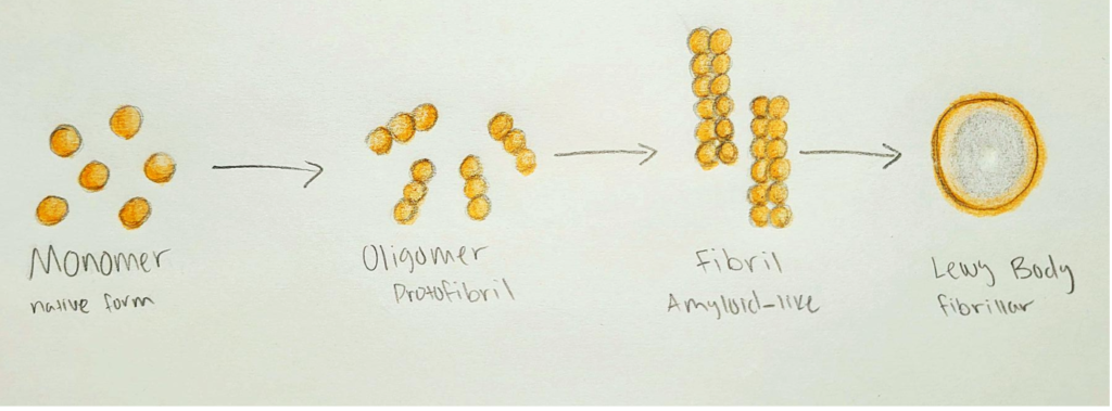
My drawing showing different alpha-synuclein conformations
The process of aggregation is believed to involve a transition from monomeric to oligomeric forms, which are thought to be more toxic to neurons. This can disrupt cellular processes, cause oxidative stress, and trigger inflammatory responses in the brain, ultimately leading to the death of dopamine-producing neurons.
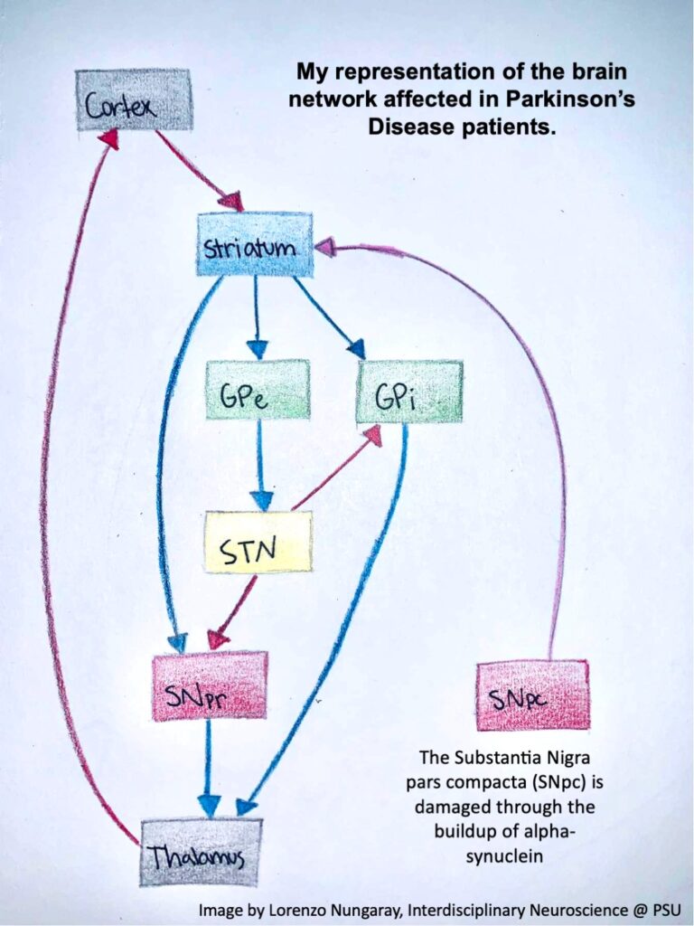
The loss of these neurons in the substantia nigra, which release a neurotransmitter called dopamine, is a hallmark of Parkinson’s disease and underlies the motor symptoms observed in patients.
There are other neurological disorders caused from the aggregation of alpha-synuclein known as synucleinopathies. These disorders involve multiple system atrophy and dementia with Lewy bodies.
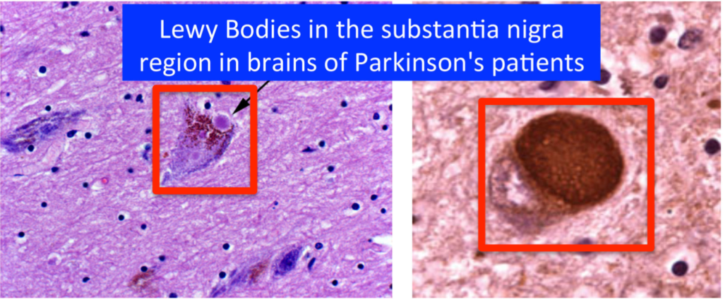
IMAGE: Journey with Parkinson’s
Sleep removes toxins
Normally, alpha synuclein buildups should be eliminated by the glymphatic system which has its most productive state during sleep. The glymphatic system expands during sleep and allows toxins, and metabolic buildup to pass through and be cleaned out of the brain.
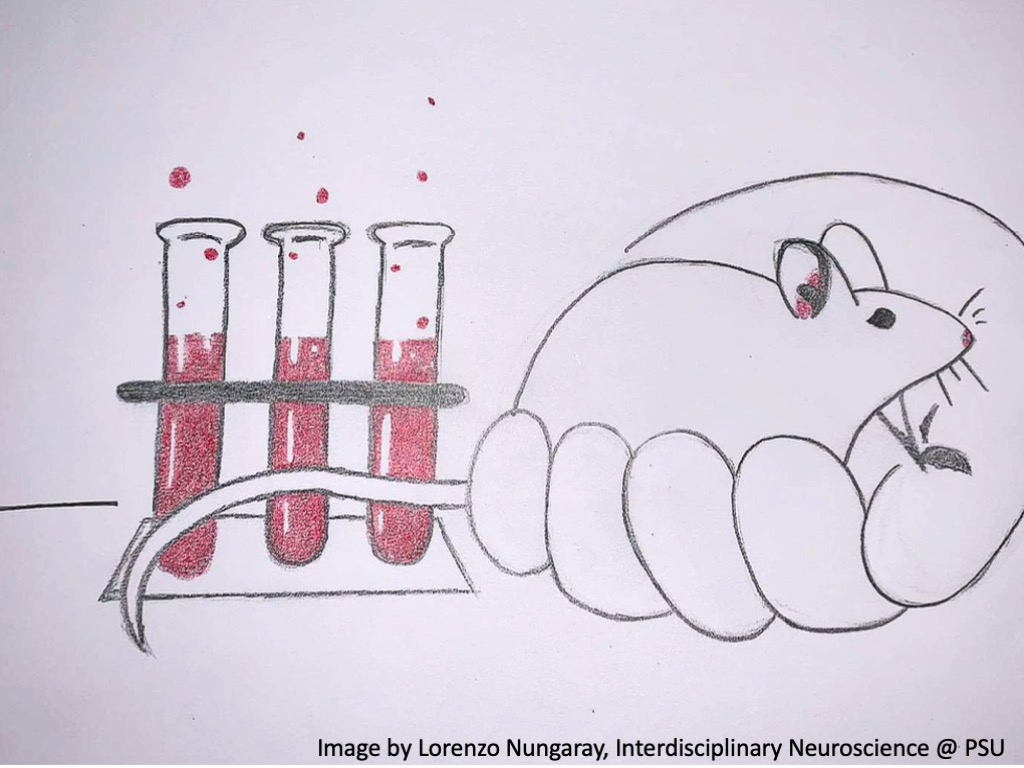
This model of Parkinson’s Disease is what the SHARP lab is focused on. We use mouse models to simulate the buildup of alpha synuclein in the brain and how this affects the behavior and physiology of the mouse. A method that is used at the SHARP lab to assess the degeneration of movement in mice with Parkinson’s Disease is tracking their gait using sensors and a treadmill.

IMAGE: Mouse on Digigait Imaging System. This picture is not property or affiliated with PVARF/OHSU
This software tracks the mouse’s movements when walking and analyzes the data collected to determine whether the mouse has an abnormal gait. An abnormal gait is one of the symptoms of Parkinson’s Disease so this method can tell us a lot about the effects of Parkinson’s on the motor function of mice.

How much sleep do you need?
This graphic shows how much sleep you should be getting a night on average. The amount of sleep needed is highly dependent on the age of the individual. Newborns should be getting around 14 to 17 hours of sleep a day while people over 65 years old on average get 7-8 hours a night. This trend of decreasing amounts of sleep persists throughout one’s life as shown on the graphic.
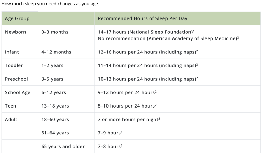
IMAGE: How Much Sleep Do I Need?
The graphic below compares the estimated amount of sleep the U.S. population has received per night starting in 1942 and ending in 2013. The trend shows that many of us get less sleep now than before.
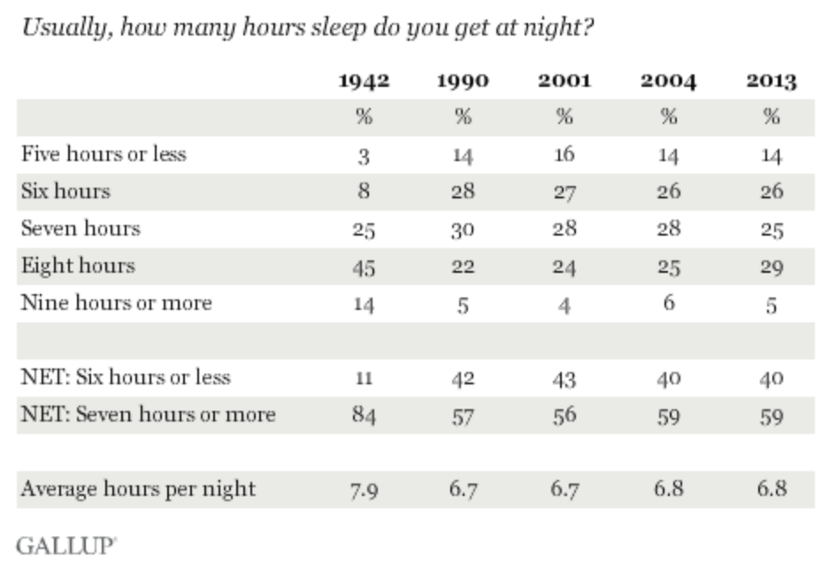
LEARN MORE: In U.S., 40% Get Less Than Recommended Amount of Sleep
The graphic shown below shows the states that on average have the most people who get fewer than 7 hours of sleep at night (red) with Hawaii being the highest percentage in this regard. The green states represent the average number of people who get over 7 hours of sleep a night with Colorado being the highest.

LEARN MORE: 100+ Sleep Statistics
As I’m sure we all know, most of the population does not get the recommended amount of sleep per night that we should be getting. What does this mean? This means that most of the population is now more susceptible to neurological diseases now than ever before!
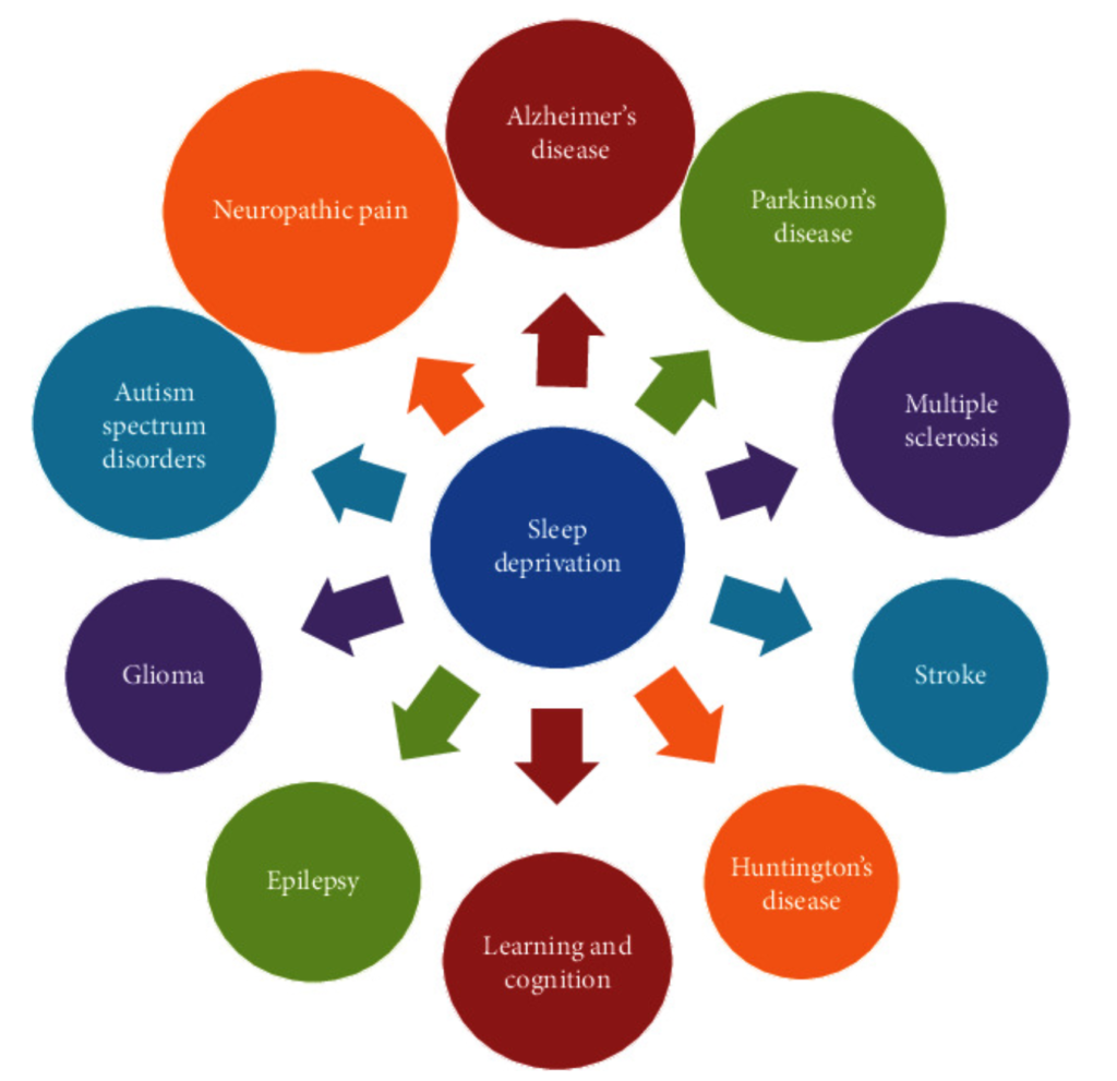
LEARN MORE: Sleep Deprivation and Neurological Disorders
Protect yourself
What other steps can we take to help prevent this? There are many factors other than sleep that can influence the onset of neurodegenerative diseases such as Parkinson’s and Alzheimer’s. Some of these factors include lifestyle choices such as diet and exercise habits. Some factors are out of our control such as genetic makeup.
Of course there are many other side effects of sleep deprivation that we still have not touched on. These can include changing in mood, memory, and overall brain function.
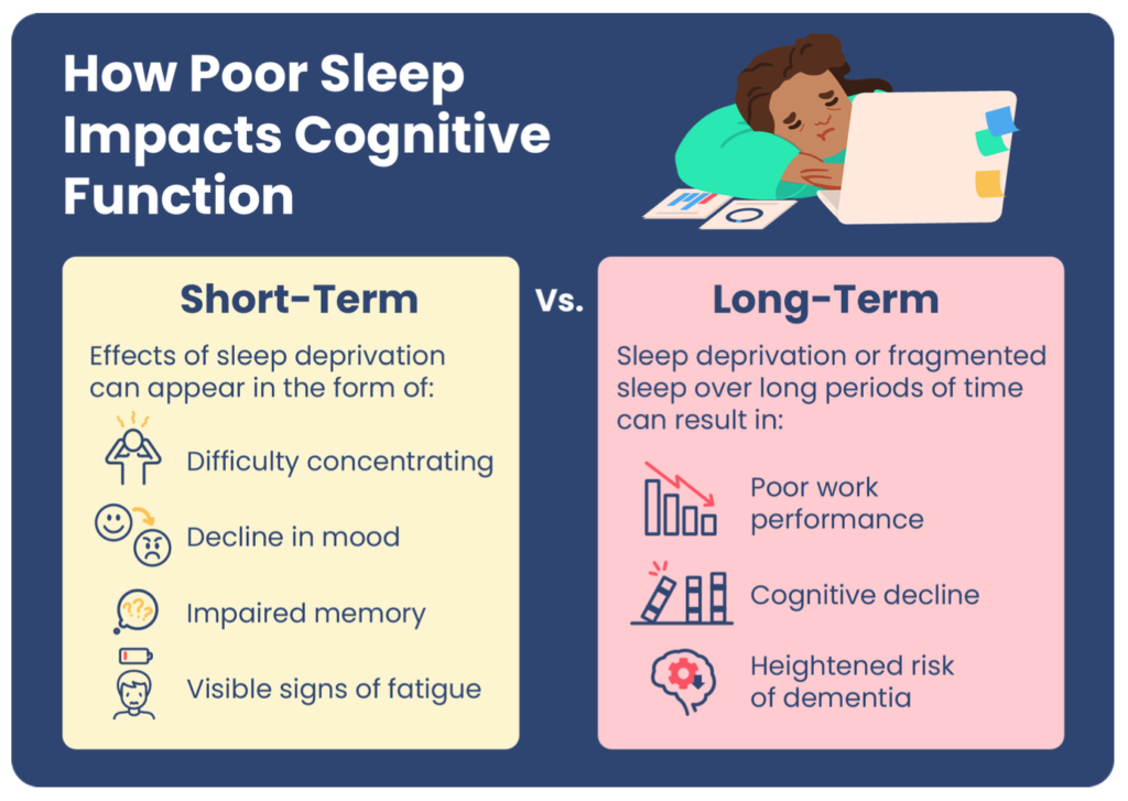
LEARN MORE: How Lack of Sleep Impacts Cognitive Performance and Focus
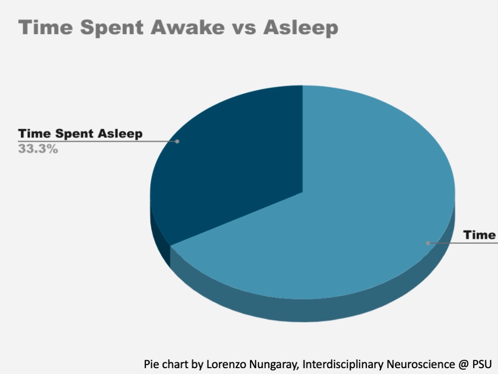
Sleep is very important and is highly conserved throughout all life! The average human spends around one-third of their life sleeping!
If there is any take away from this post I hope it is that sleep is very important to one’s health. Sleep should be taken seriously by everyone at every age!
Special thanks to SHARP for giving me the opportunity to work with them and be a part of the great work they do. I really appreciate being able to work and learn alongside the amazing people working at the SHARP lab and hope to continue to work alongside them for a long time!
LEARN MORE: Baseline characteristics of the North American prodromal Synucleinopathy cohort
LEARN MORE: Amyloid-β Dynamics Are Regulated by Orexin and the Sleep-Wake Cycle
LEARN MORE: Brain may flush out toxins during sleep
LEARN MORE: Mouse-brain model maps spread of alpha-synuclein in Parkinson’s disease


