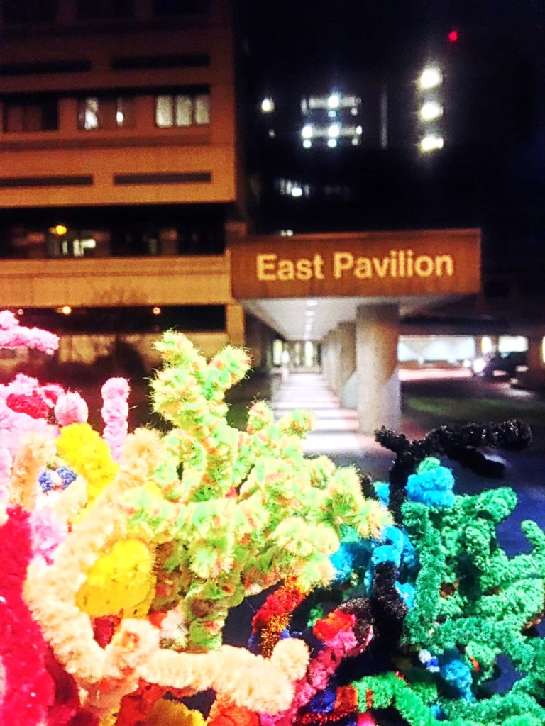
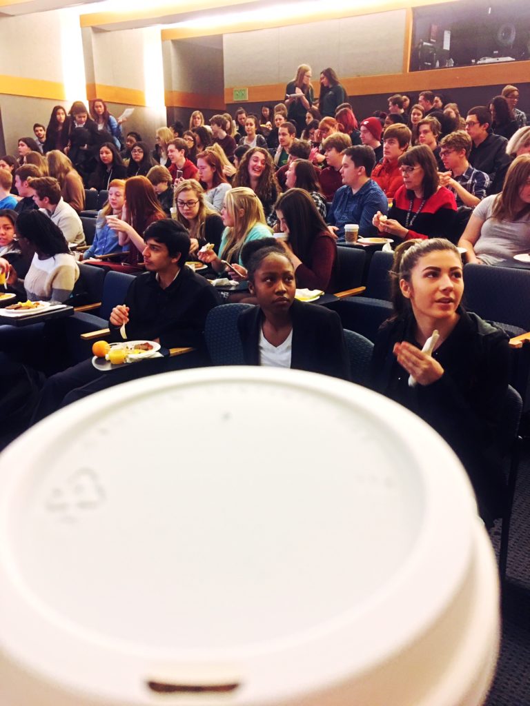
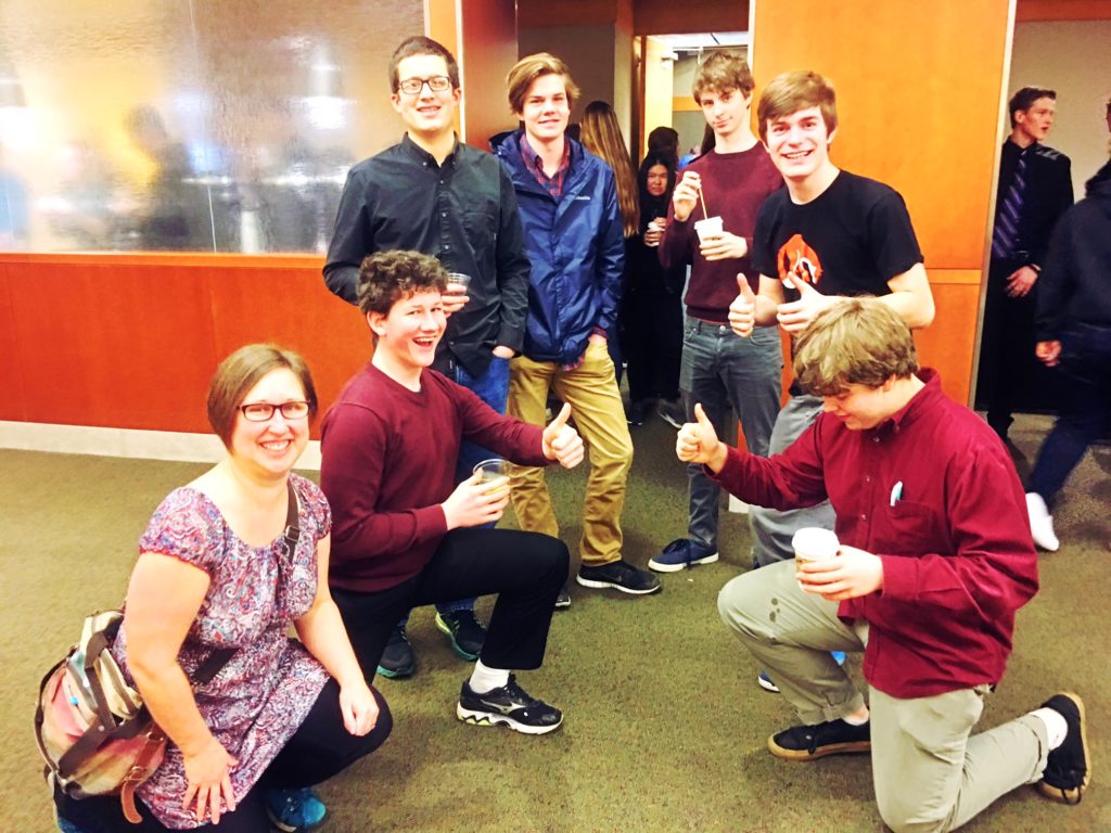
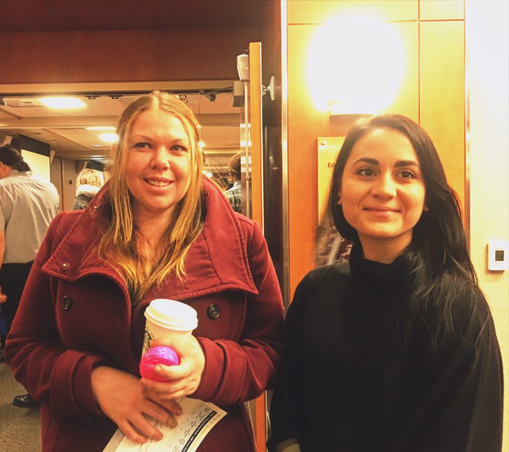
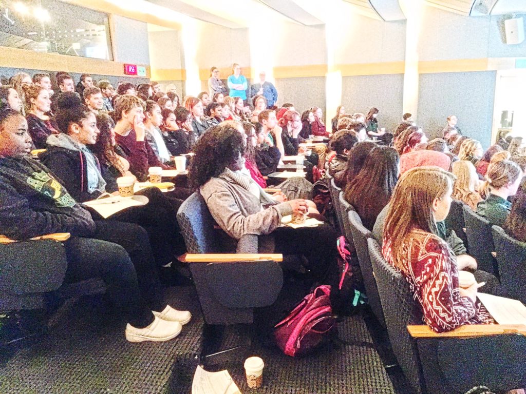
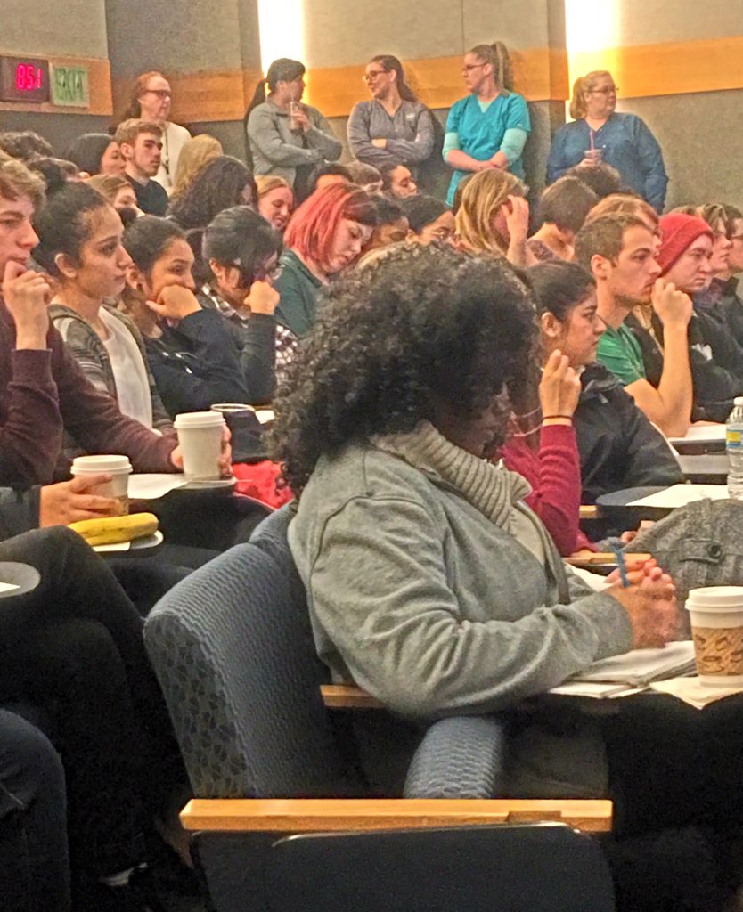
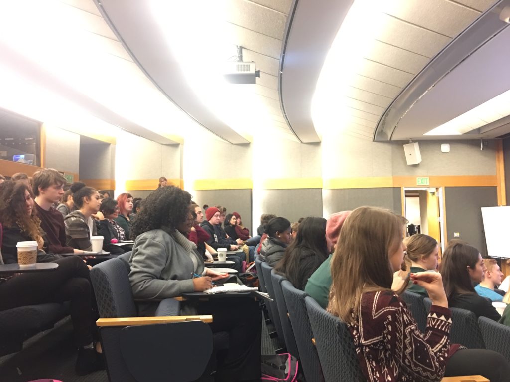
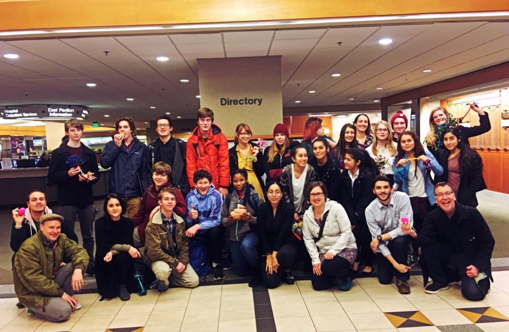
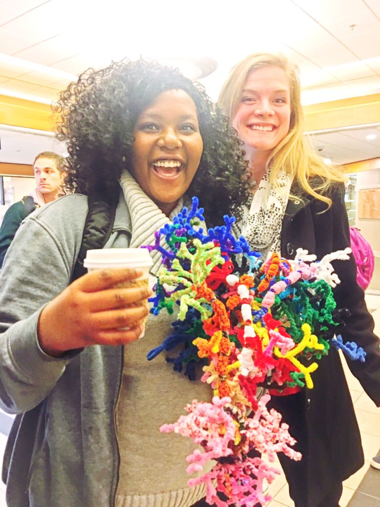
Noggin was honored to bring 30 high school students, teachers and our own volunteers to Providence St. Vincent hospital this morning, to participate in the Providence School Outreach Brain Watch program, and view an actual surgery on a living patient’s brain..!
We arrived EARLY, around 6:15am, and major props to all the young people from Jefferson High School, Benson High School, Madison High School (in Portland Public Schools) and Fort Vancouver High School (in Vancouver Public Schools), who tackled the dawn trek to Souther Auditorium!
LEARN MORE: Sleep in Adolescents: The Perfect Storm
Breakfast (including coffee!) provided by the Brain Watch program was welcomed by students, teachers, and our enthusiastic NW Noggin volunteers, including Jacob Schoen, Sarah Fitzsimmons, Kayla Townsley, and Heather Hamilton from Portland State University, and Joey Seuferling, Bradley Allen Mahoney, Sterling Gray and Kim Engeln from WSU Vancouver!
LEARN MORE: Arousal effect of caffeine depends on adenosine A2A receptors in the shell of the nucleus accumbens
Kayla and Heather are also participants in the NIH-funded BUILD EXITO, an undergraduate training program that arranges advising and mentoring support for students interested in biomedical, behavioral, clinical, health and social science research careers…
LEARN MORE: BUILD EXITO
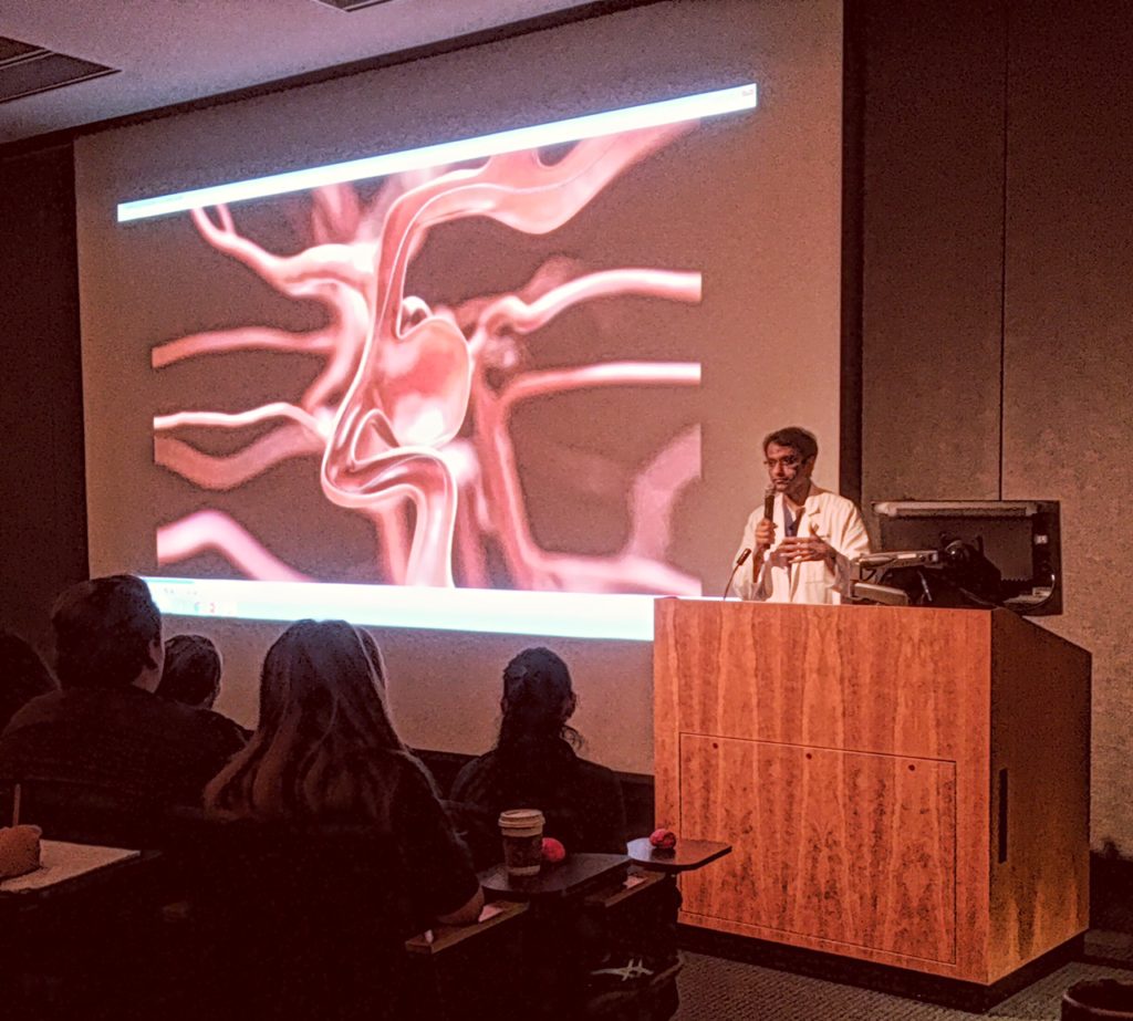
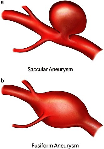
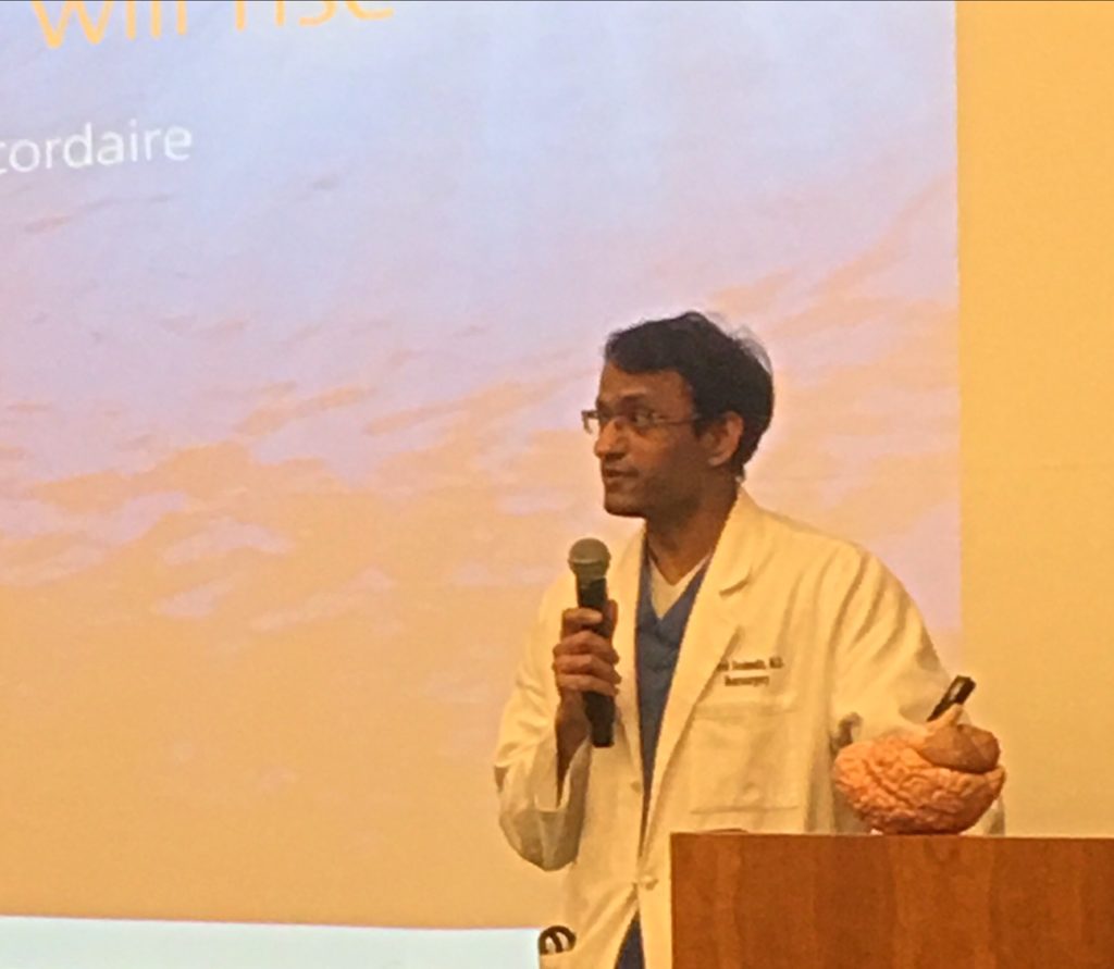
Everyone woke up as we heard from a variety of clinical professionals, including the neurosurgeon, Dr. Vivek Deshmukh, a skilled and soft-spoken expert who completed his extensive medical training at age 33 (!). Dr. Deshmukh performed last year’s Brain Watch surgery, which also involved the clipping of a bulging weak spot in a blood vessel wall, known as an aneurysm…
LEARN MORE: Brain Watch Wednesday!
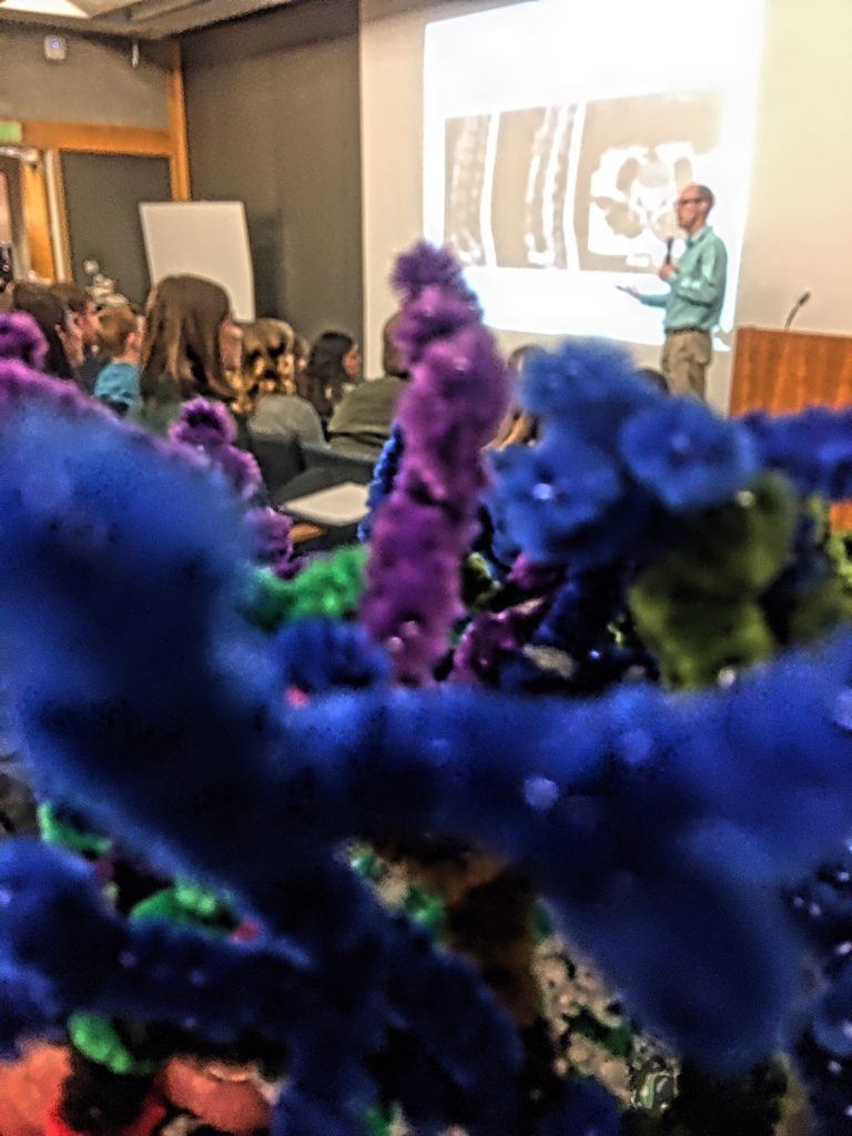
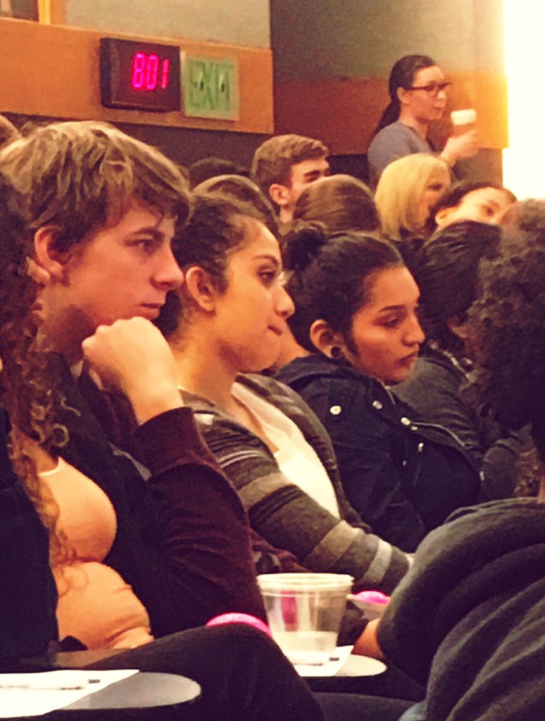
Our students were fascinated to hear about his patient’s case, and also learn from other medical professionals, including a nurse practitioner, an MRI technologist, and a physician’s assistant.
We learned how aneurysms, those balloon like out-pockets of weakened vasculature, are capable of bursting, which can result in a devastating bleed – a hemorrhagic stroke, loss of brain tissue and even death. It’s estimated that 1 – 5% of us has an aneurysm, typically at a branch point where blood pressure is most intense. Aneurysms take years to develop, and most people have no idea they’re present unless they burst…
LEARN MORE: Prevalence of unruptured intracranial aneurysms
LEARN MORE: Hemorrhagic stroke
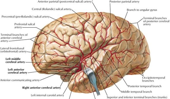
This particular patient’s aneurysm was discovered, as many are, from an imaging scan taken for a different medical reason. But her abnormality was large, and in a dangerous branching spot on the left middle cerebral artery (MCA), a critical blood vessel which emerges from deep in the brain through the Sylvian or lateral fissure and feeds important cortical regions in the left hemisphere, including many associated with language comprehension and speech…
Dr. Deshmukh would need to delve deeply into the Sylvian fissure, carefully clipping his way in using tiny scissors and additional dissection tools, avoiding insult to brain tissue and blood vessels, until he reached the dangerous bulge towards the middle of the patient’s brain…
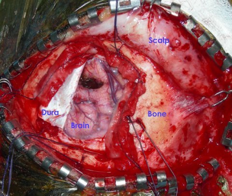
However, first he had to gain access to the brain, and as the screen filled with images of the patient’s scalp pulled back, her temporalis muscle cut, and we heard the whine of the craniotome (the special saw used to cut through bone), we were riveted and silent ourselves…
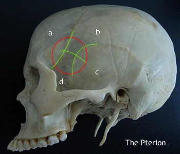
This year’s craniotomy was pterional, which meant that Dr. Deshmukh cut the cranium (skull) near the pterion, the region where several skull bones, including the frontal, parietal, temporal, and sphenoid join together. It is located on the side of the skull, just behind the temple…
LEARN MORE: Pterional craniotomy
(NOTE: NOT FROM TODAY’S SURGERY)
Once the bone was removed, wrapped in an antibiotic-soaked sponge and transferred to a sterile tray, Dr. Deshmukh took his seat in a special chair with supportive armrests designed to stabilize his hands. He operated a camera with his mouth, and we were treated to the same exact images he viewed as he cut through the meninges, the outer layers protecting the fragile brain…
IMAGE SOURCE (NOTE: NOT FROM TODAY’S SURGERY)
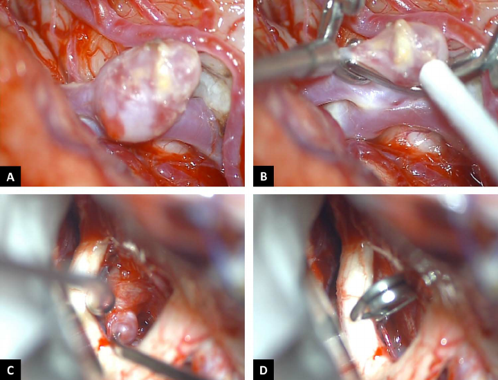
The surgeon continued to carefully cut his way through the thin, spidery-gray fibrils of the arachnoid, and then worked his way deeper and deeper into the lateral fissure. When we actually saw the aneurysm, several of us gasped, as it looked large under the microscope, with several reddish bulges indicating the weakest spots most likely to blow…
IMAGE SOURCE: Intracranial aneurysm arising from infundibular dilation
(NOTE: NOT FROM TODAY’S SURGERY)
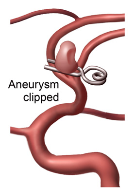
With exquisite precision, Dr. Deshmukh gently isolated the aneurysm, and then opened the titanium clip used to permanently seal it off from the MCA. This was the most intense moment of surgery, as he used thrombin-infused sponges to mop up some limited bleeding, and decided on the precise spot for clip release…
To confirm a successful clip placement, a fluorescent dye was injected into the patient’s bloodstream, the lights were turned off, and the channeled flow of blood was revealed in a bright whitish-blue flow through vessels, with no fluorescence detected in the now isolated aneurysm bulge!
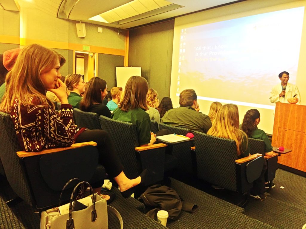
As the physicians assistant began closing up, Dr. Deshmukh joined us in the Souther Auditorium to take questions from students clearly fascinated and energized by what they’d seen. We donated our Noggin brain model to the podium, as our surgeon went over details of the procedure, discussed his educational background and career, and offered advice for those interested in working in the demanding and exhilarating field of healthcare…
An extraordinary educational morning! Our great thanks to Toyin Oyemaja and Allie Bieker of the School Outreach Program at Providence St. Vincent for welcoming us back to Brain Watch this year!
LEARN MORE: Register for FREE Career Highlights Classes at Providence!


