")
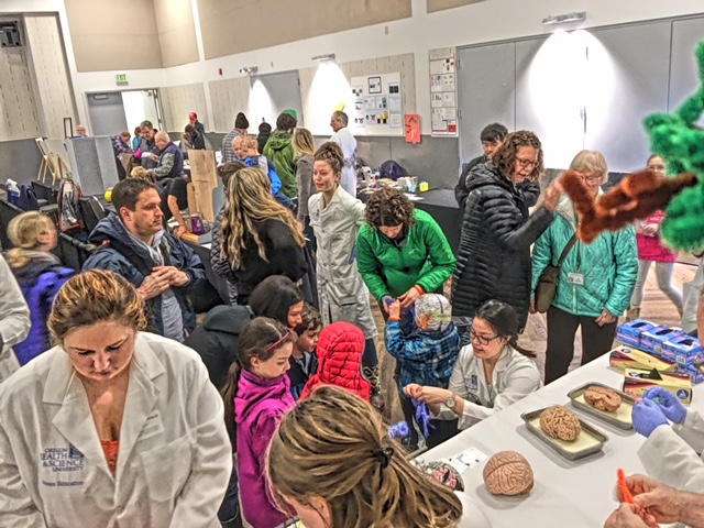
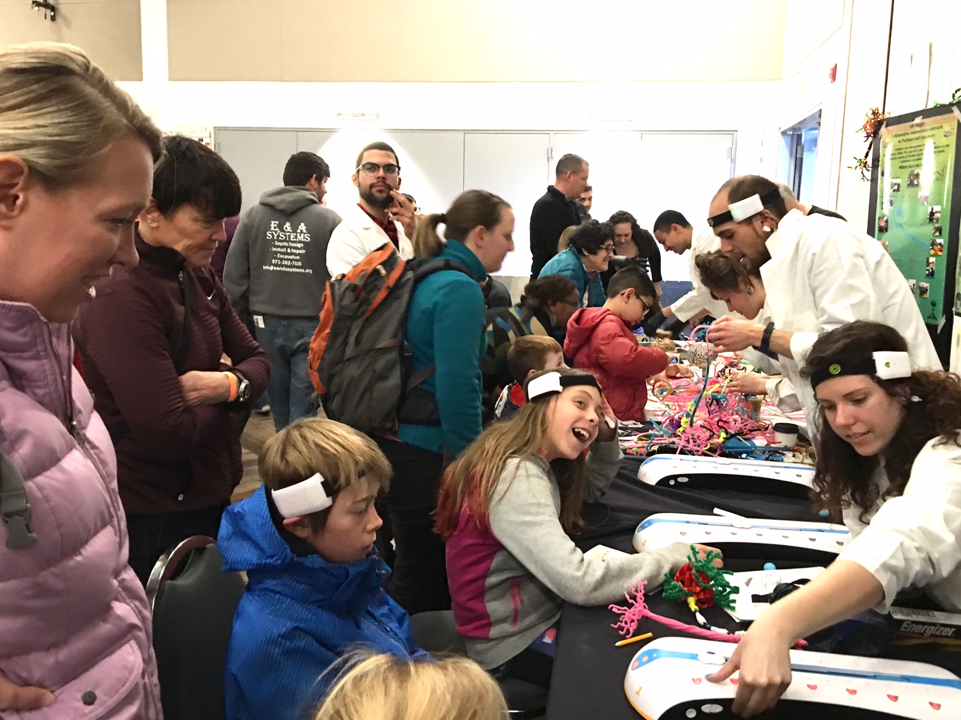
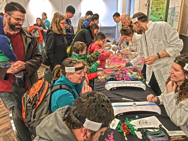
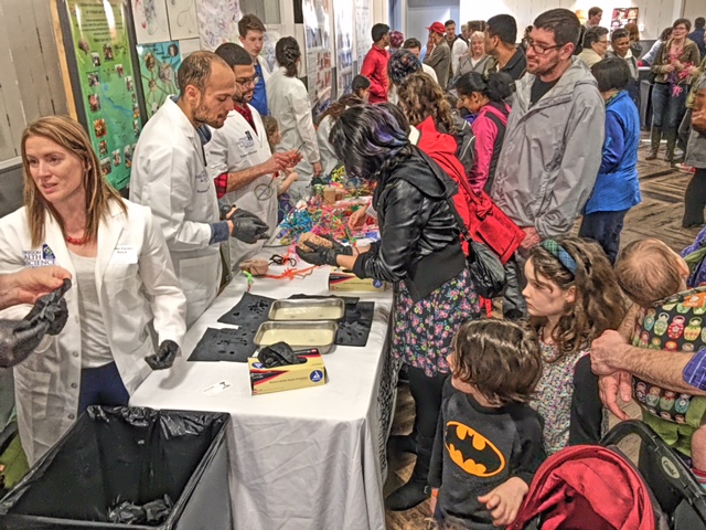
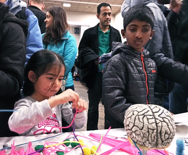
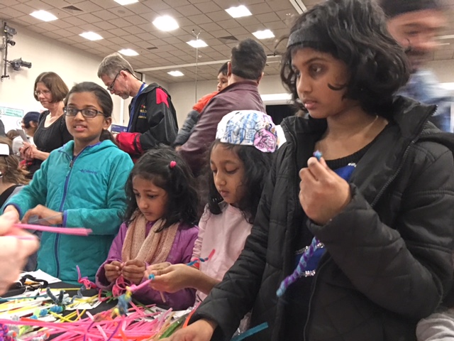
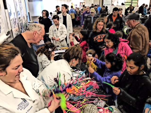
Our subcortical sensory nuclei were nearly overwhelmed by masses of eager children, parents, grandparents and members of the public curious about neurons at the annual OHSU Brain Fair!
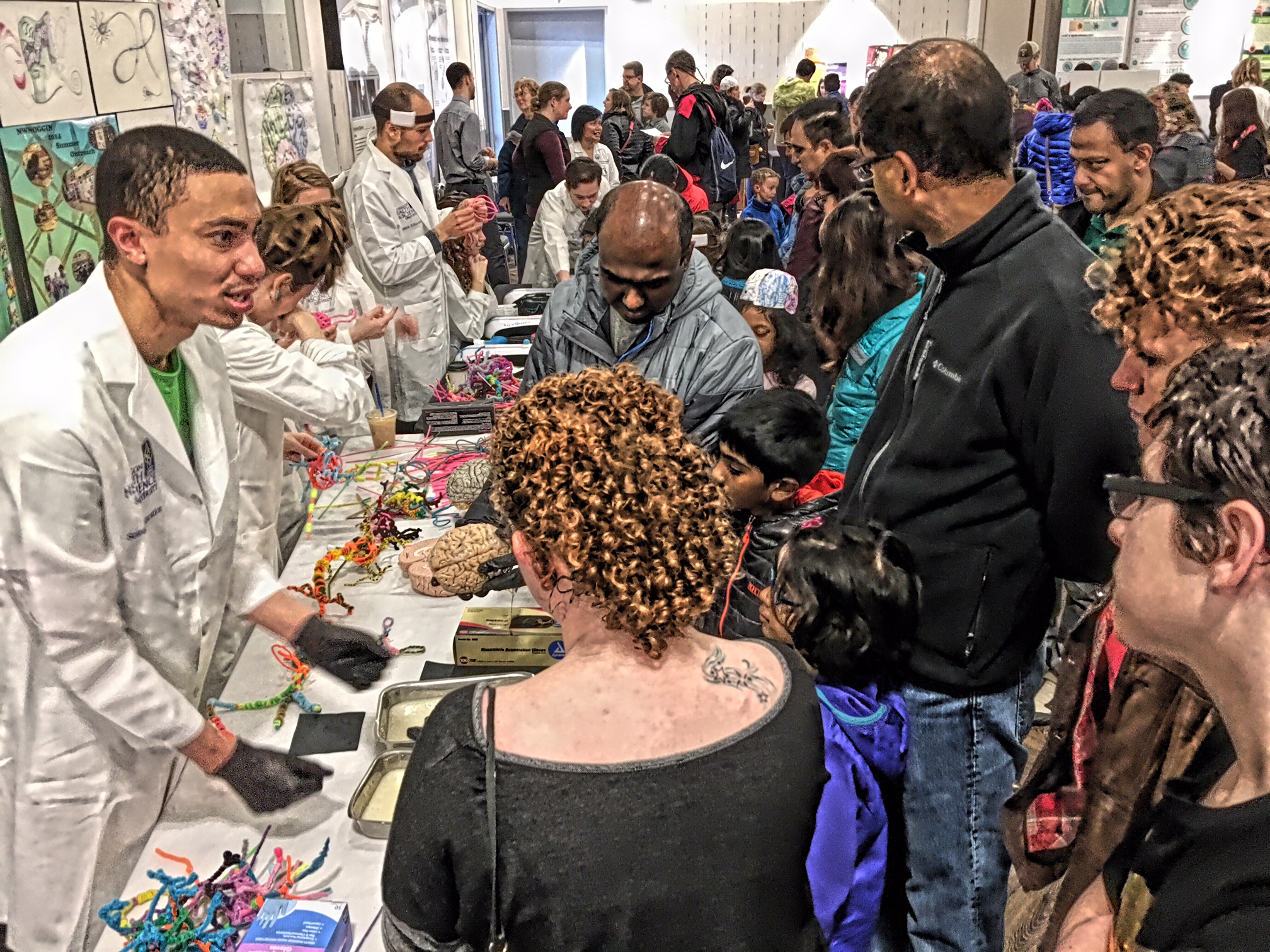
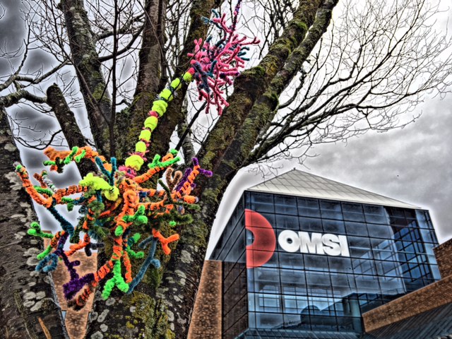
Held at the Oregon Museum of Science & Industry (OMSI), this exceptionally popular event brings together scientists, medical professionals and educators, and is the largest brain-related public celebration in the country…
The thalamus is an important collection of brain cells found right in the middle of our brains, just above the brainstem, and it is composed of separate clumps of neurons (called nuclei) that each process a specific sensory input, including vision, hearing, balance, touch, pain and taste.
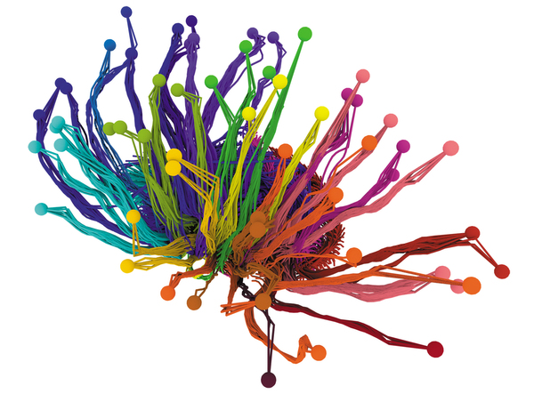
Thalamic nuclei send and receive information to and from specific areas of cortex, as illustrated in this striking image above, from the Allen Institute for Brain Science. The thalamus is that kidney-shaped structure in the middle of the picture, and those dots are connected areas in the wrinkled cortex in various lobes of the brain…
EXPLORE MORE: The role of the thalamus in the flow of information to the cortex
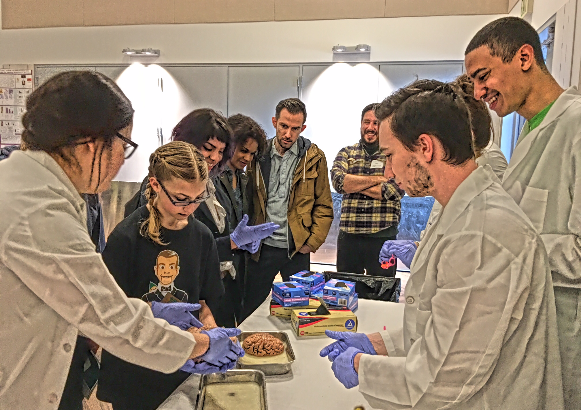
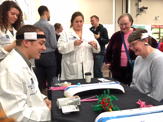
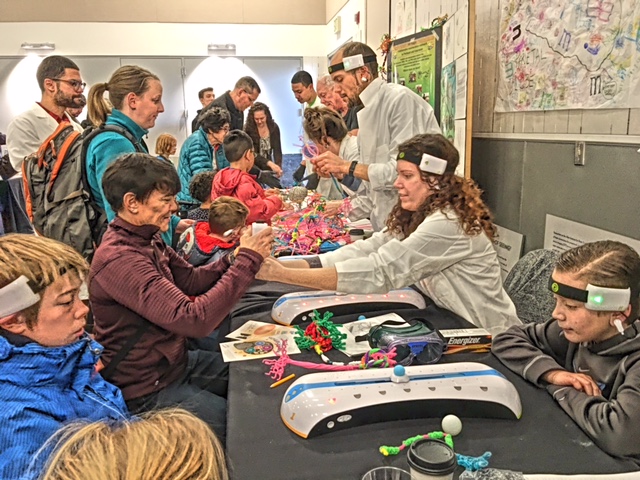
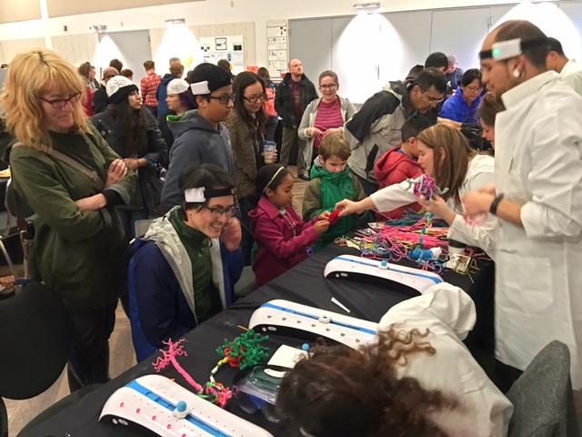
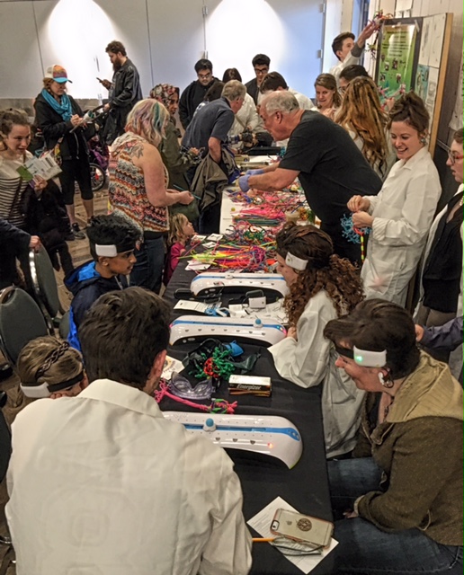
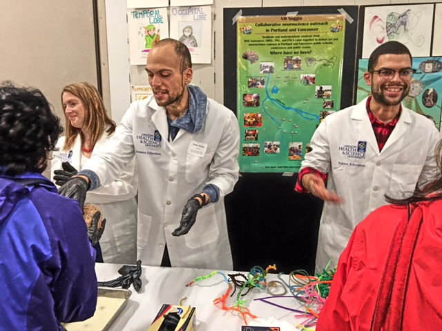
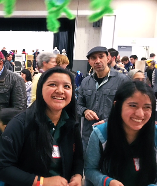
There was certainly a thunderous amount of sensory stimulation to process at OMSI! However our huge number of knowledgeable Noggin volunteers, from OHSU, WSU Vancouver and Portland State, ably described the structure and function of the thalamus, along with other brain regions, at this well-attended event…
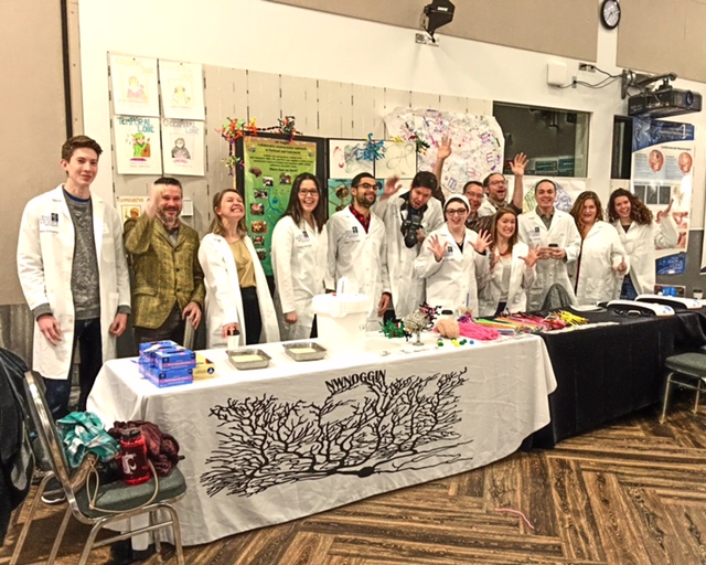
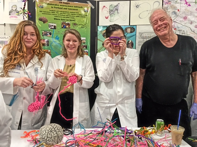
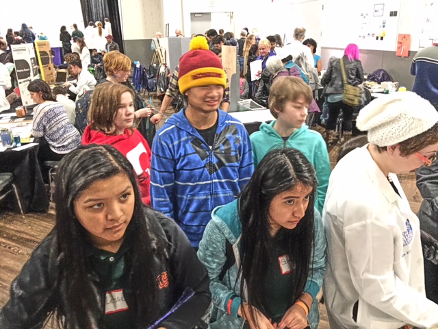
This was just part of the morning crew – and several volunteers stayed all day, from 9:30am – 5:00pm!
Our tireless graduates and undergraduates included Andre Walcott, Vanessa Jimenez, and Erika Cuellar from OHSU, Benjamin Buck, Eloise Forest, Aldair Rodriquez, Julian Rodriquez, Alex Voigt, Jessica Patching-Bunch, Allie Clark, Katherine Hill, Jacob Schoen, Jason Koch, James Reling, Austin Howard, and Jedidiah Acott from PSU, Michael White, Angela Gonzalez, Ben Yefimov, Courtney Miskell, and Chelsey Taylor Anderson from WSU Vancouver, and Laura Hogan from Marylhurst University..!
We had so many volunteers, in fact, that we sent some over to help explain the actions and effects of alcohol to OMSI visitors at the table for the Portland Alcohol Research Center (PARC) at OHSU! The PARC also just awarded $3000 to support OHSU participation in NW Noggin activities this summer!
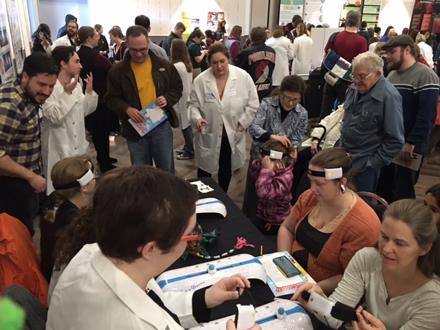
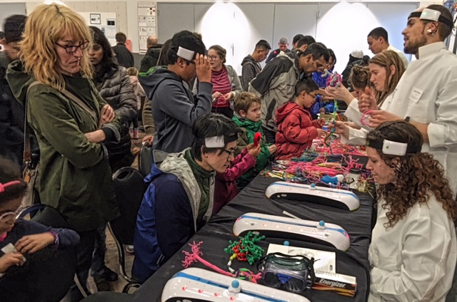
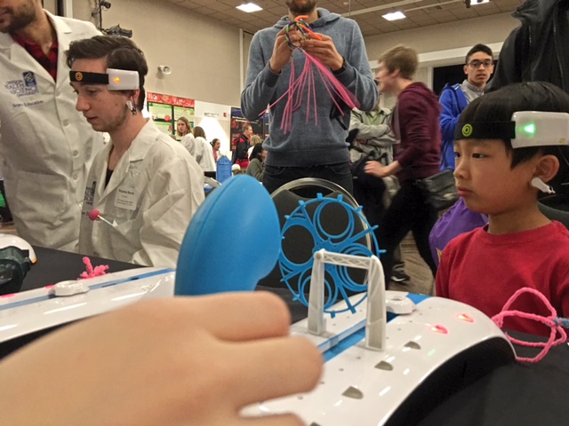
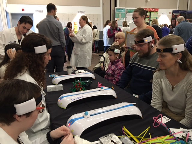
Our volunteers played Mindflex with OMSI visitors, a popular game that involves placement of a simple headband electrode over frontal cortex, and a reference electrode on the ear. That EEG signal is transmitted wirelessly to a base station, and changes in the signal – dependent on changes in what or how you think – can move a ball on a track. The goal is to mentally force that ball to your opponent’s side!
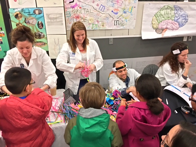
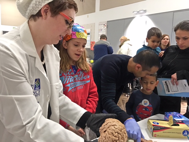
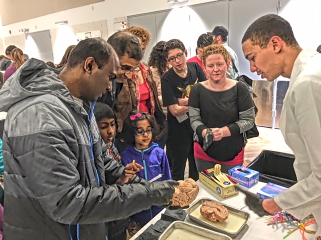
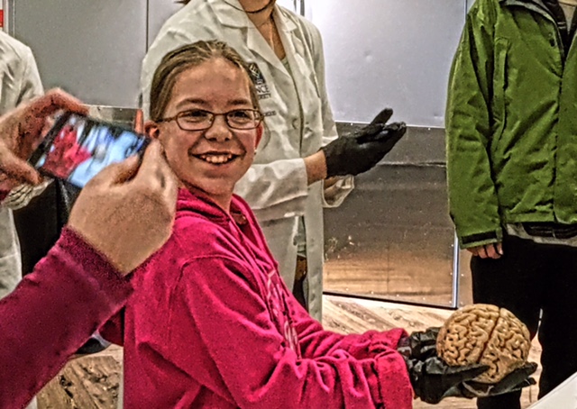
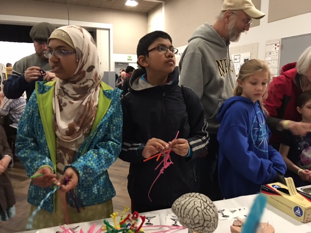
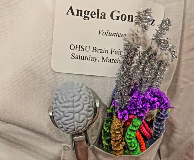
The brains were also a major draw..!
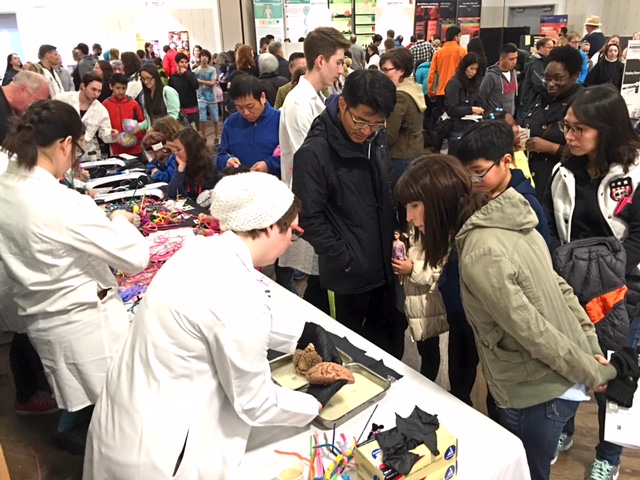
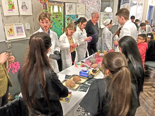
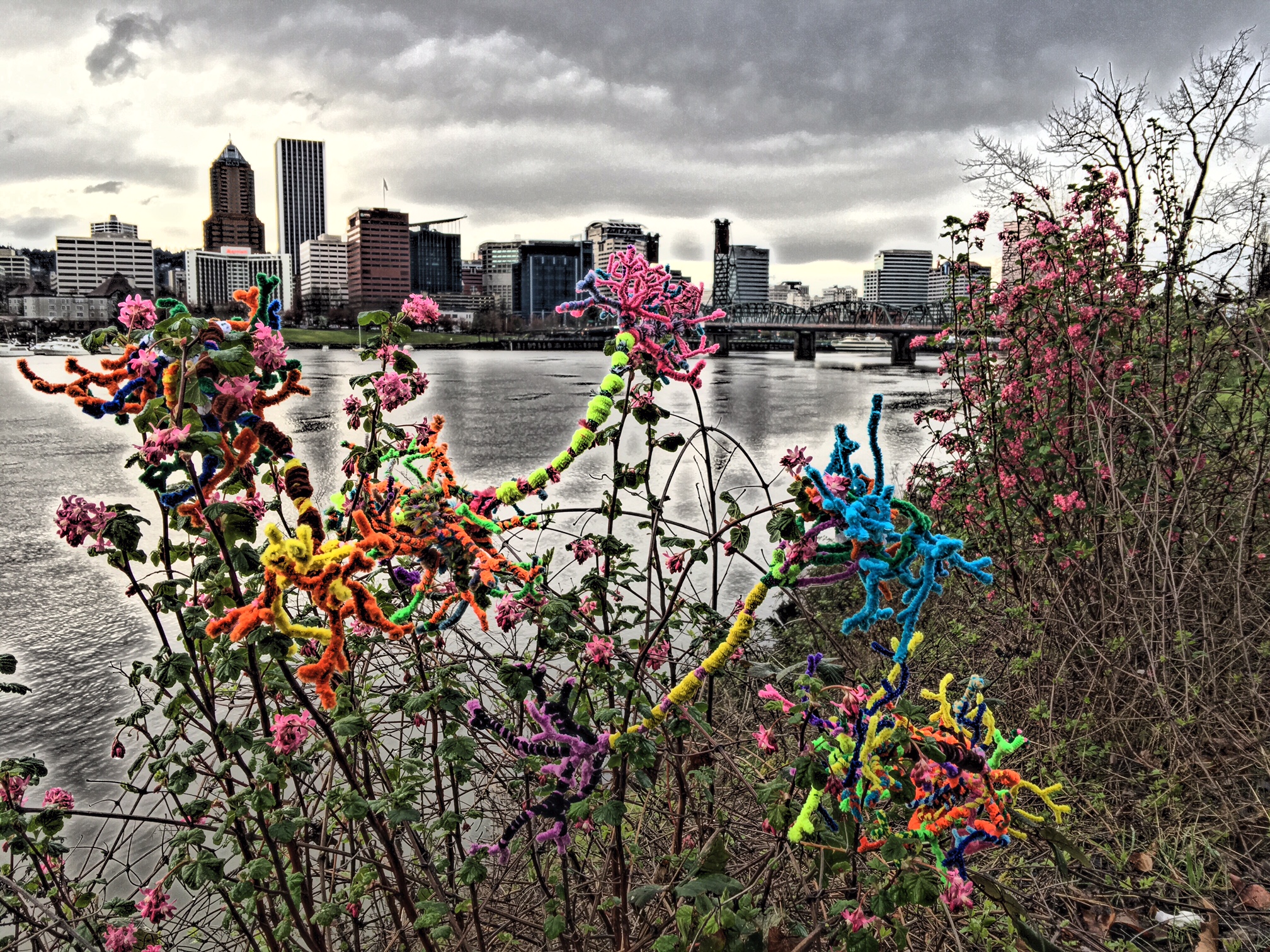
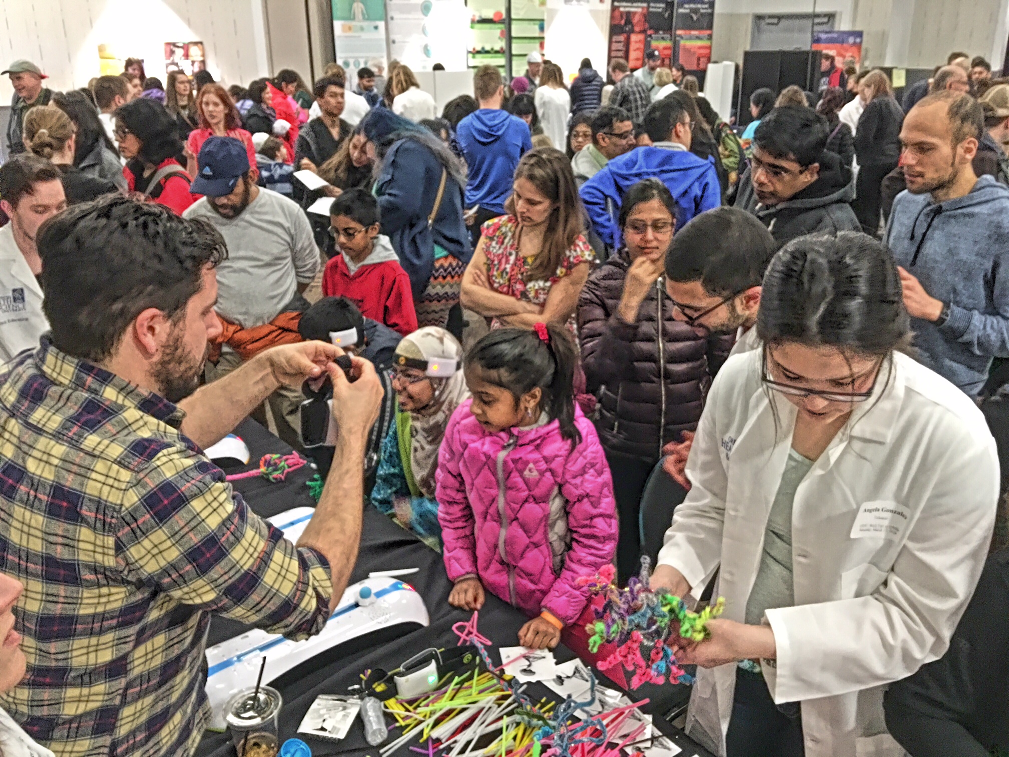
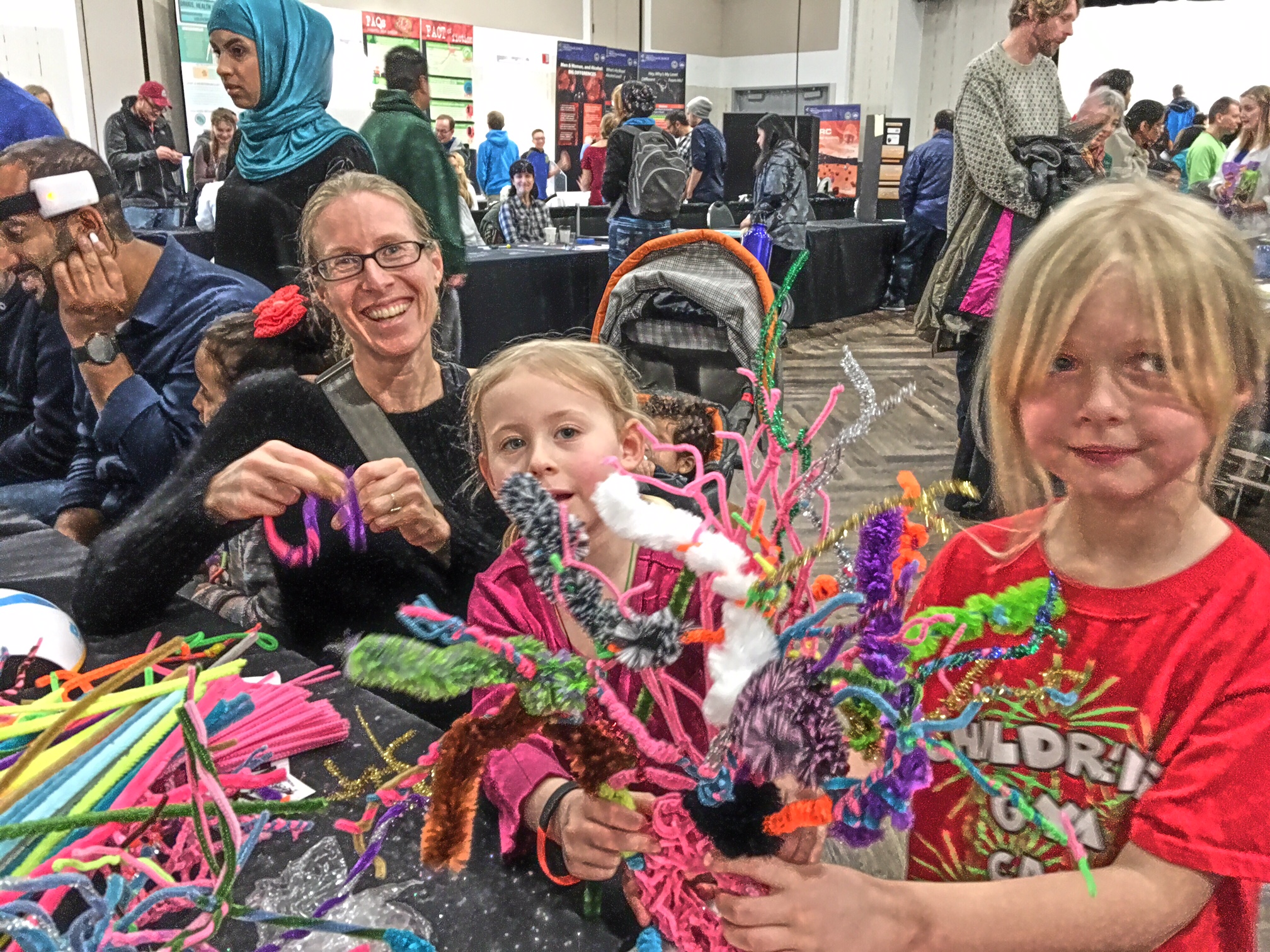
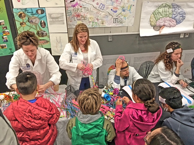
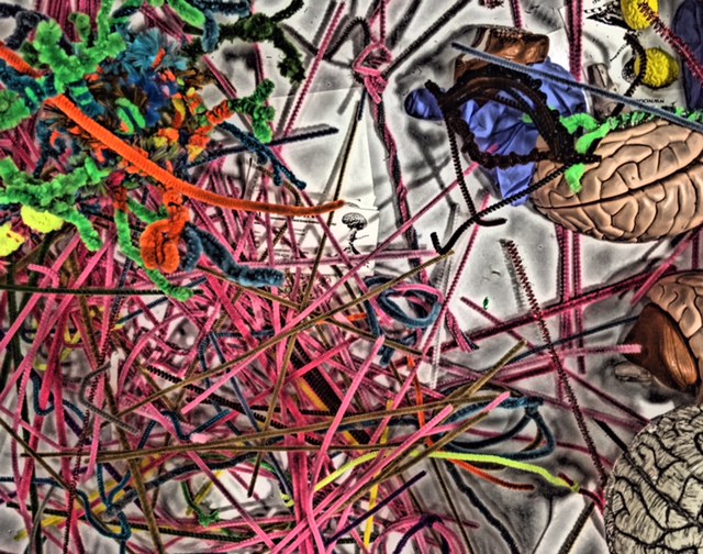
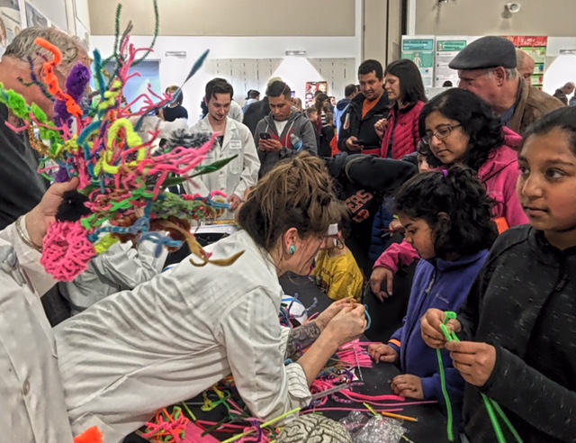
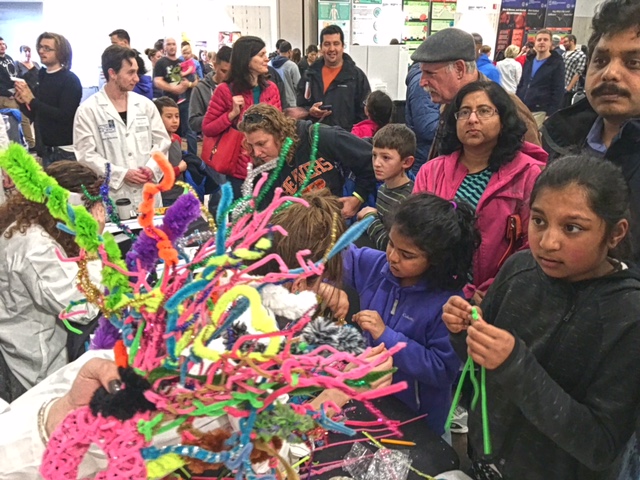
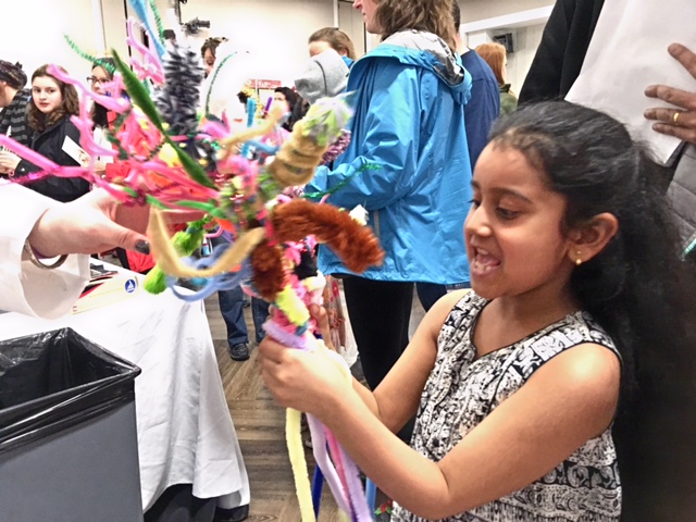
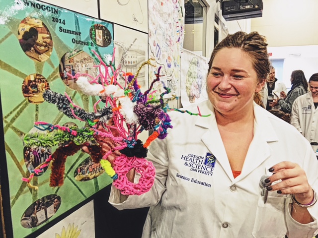
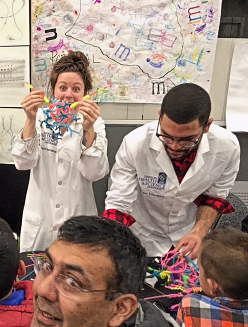
However, one of the most clearly engaging activities was neuron building – kids, adults, and many of our volunteers spent much of the day twisting pipe cleaners into colorful dendrites, axons and myelin sheaths!
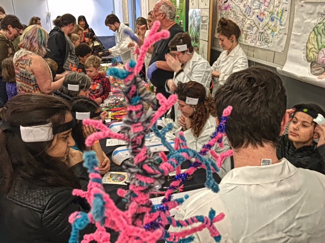
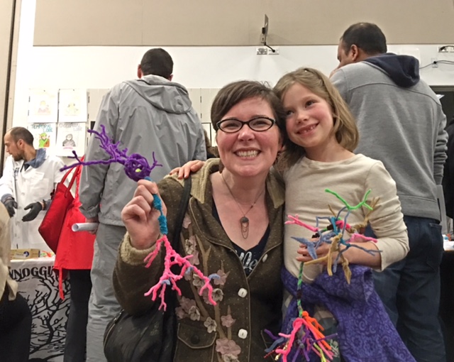
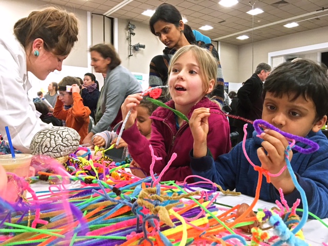
There is something particularly compelling and enjoyable about creating these cells, which are notably diverse in structure and function, so that anything you make likely has a physical analog among the billions of neurons in your brain…
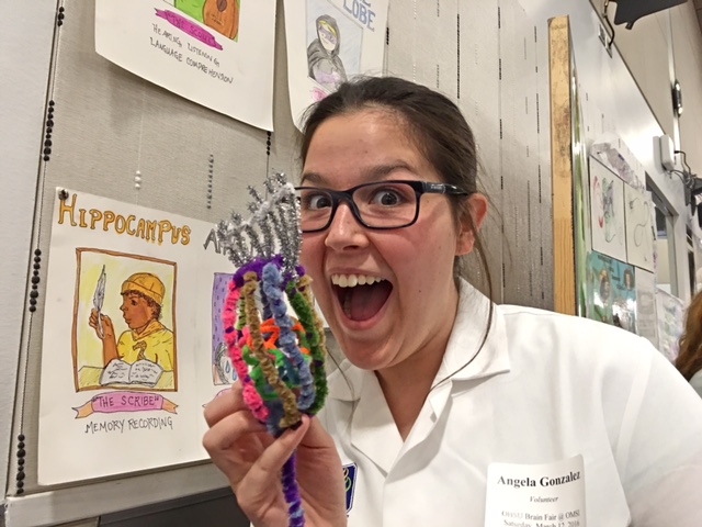
Some of our volunteers created particular neurons, and used them to explain how their unique structure determines their specialized functions. Angela Gonzalez from WSU Vancouver fabricated a hair cell, from the inner ear, with sparkling stereocilia that wave back and forth in response to sounds of different frequencies…
And Courtney Miskell, also from WSUV, constructed a dramatic Purkinje cell from the cerebellum, with extensive waving dendrites capable, in the brain, of collecting synaptic inputs from up to 200,000 sources! These cells are among the largest we have, and their axonal outputs contribute tremendously to our coordinated movements…
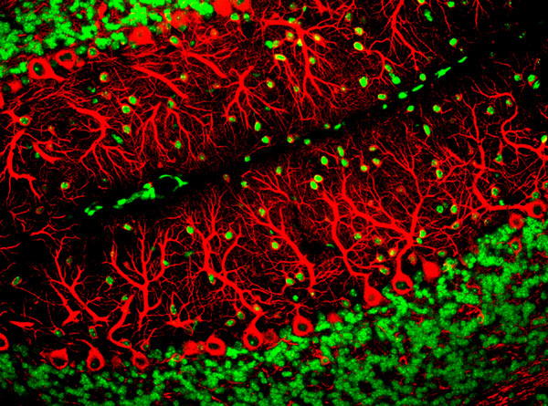
EXPLORE MORE: Purkinje cells (red) are one of the main cell types in the brain
An extraordinary day of thalamo-cortical exercising public outreach!

Many thanks to the OHSU Brain Institute for welcoming us back to OMSI – and for feeding us, too (thank you Susan Sugarman ?)!