")
Post by Joseph McKinney, senior undergraduate at Portland State University, finishing his Bachelor of Science in Science, with minors in Interdisciplinary Neuroscience, Biology, and Philosophy.

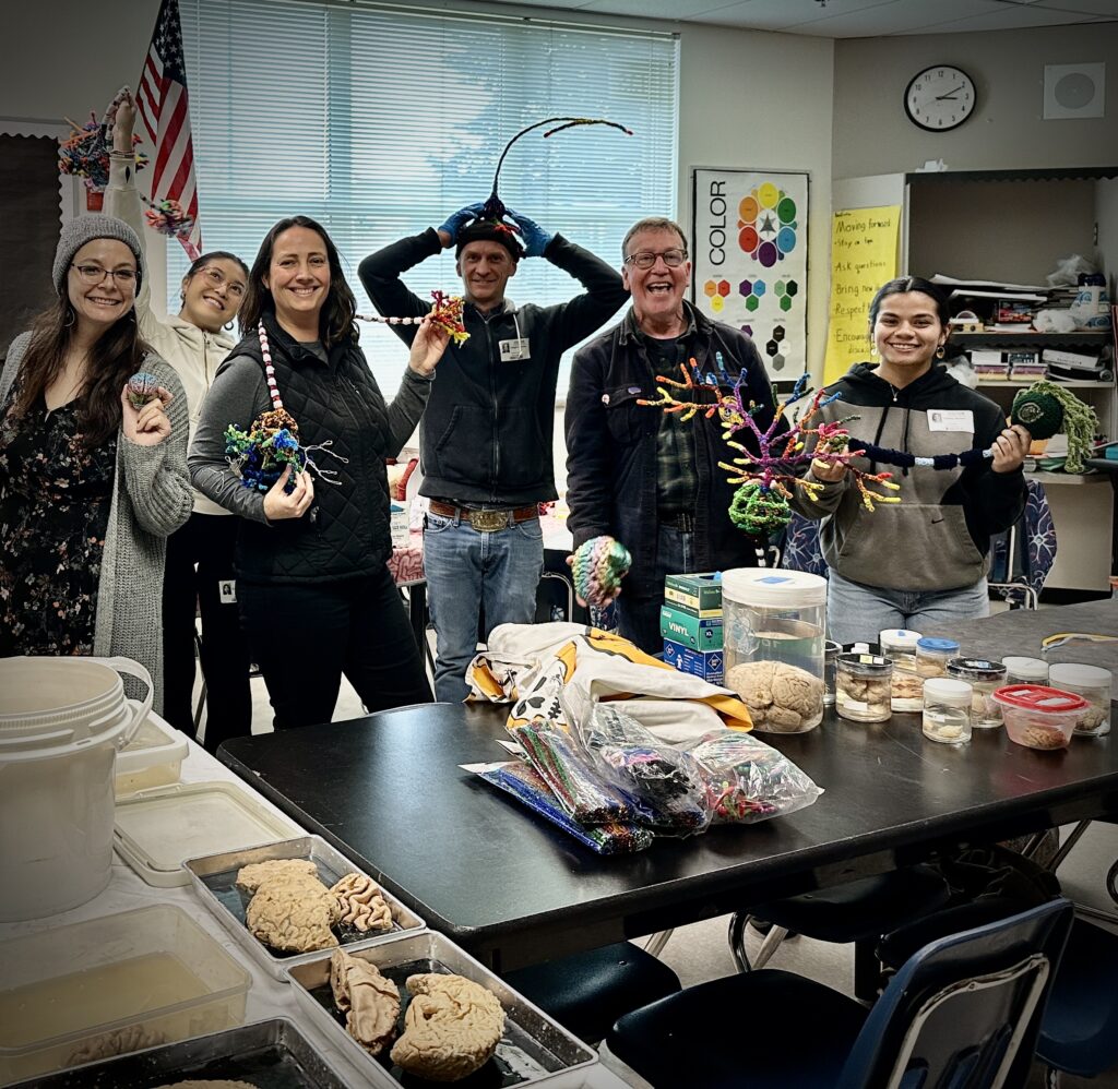
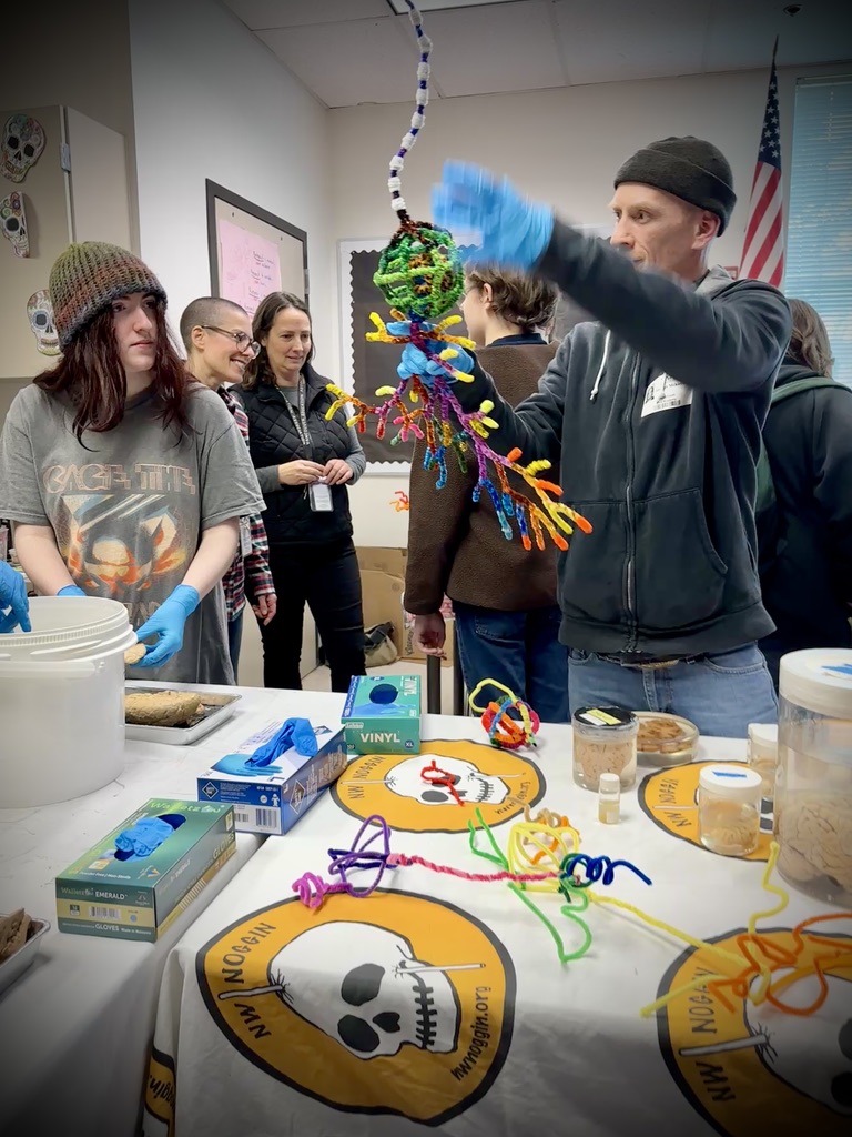
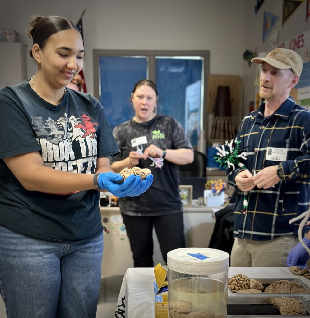
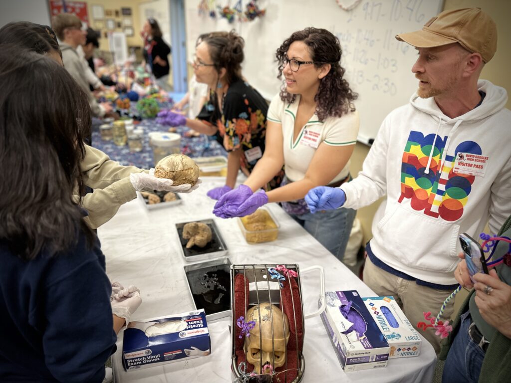
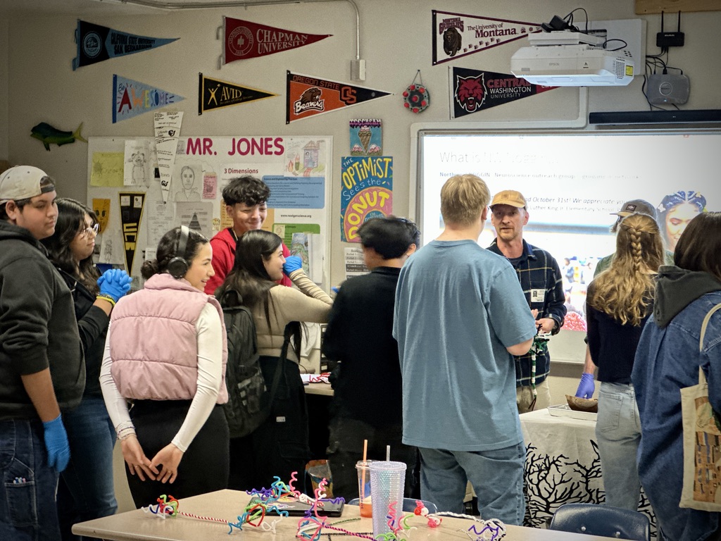
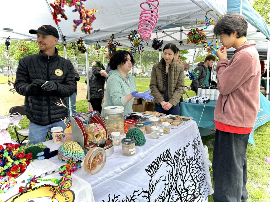
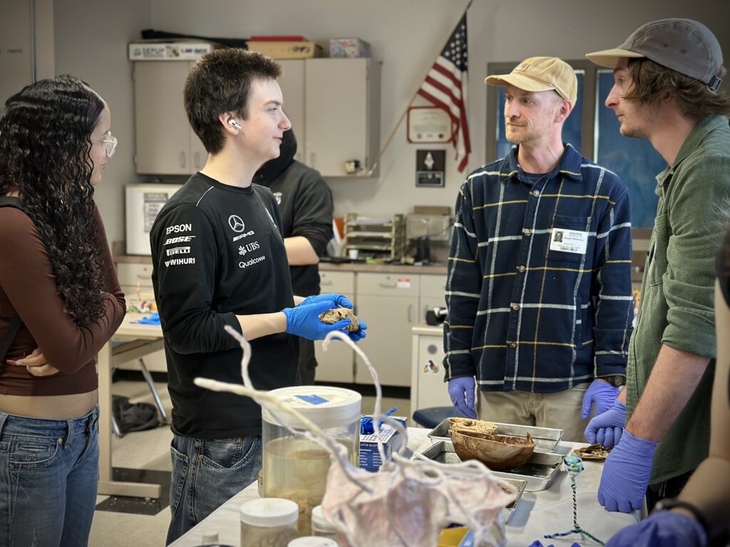
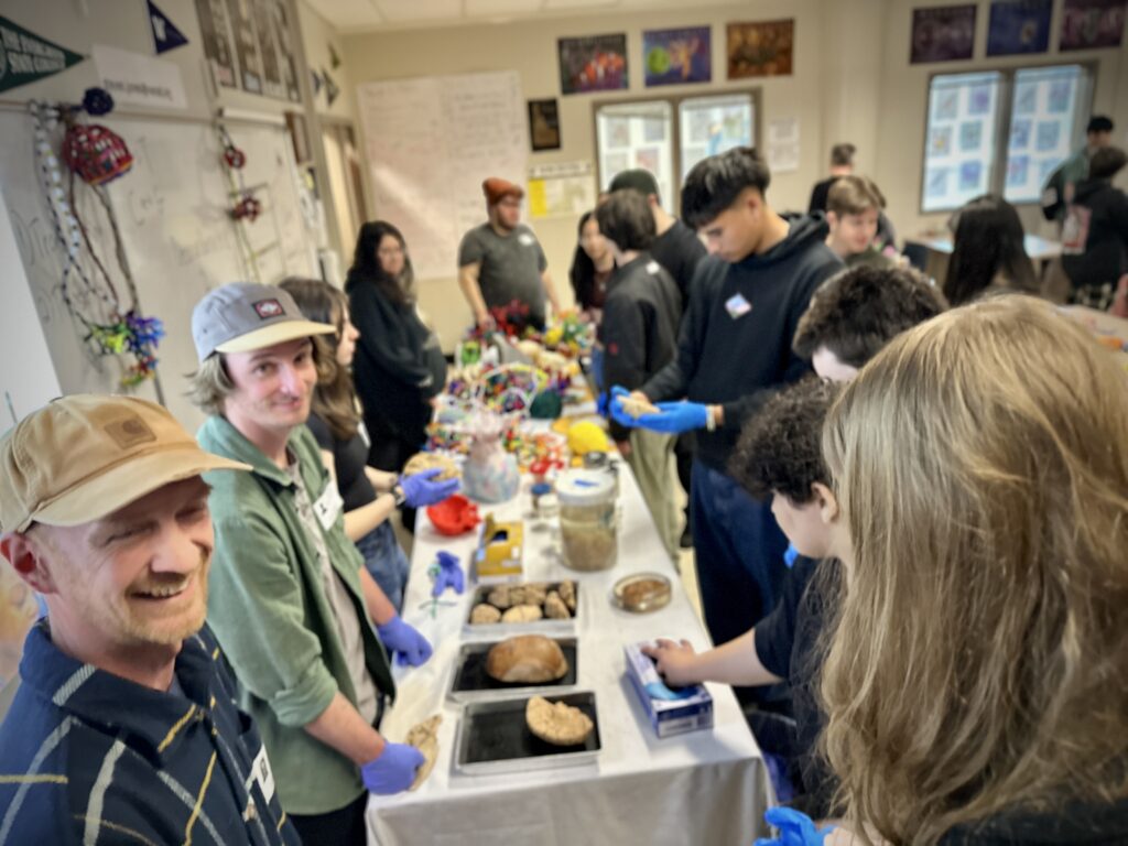
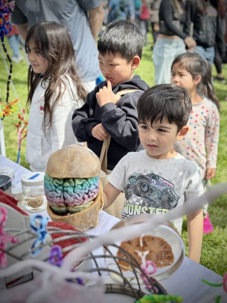
During my Winter and Spring terms at PSU, I got to join NWNoggin on some of their neuroscience and art outreach visits to different public schools around Portland and Vancouver, as well as the MacLaren Youth Correctional Facility, and local community events like New Years in the Park.
We got to bring real brains to the people, connect with students and community members over the experience, and it gave me the opportunity to learn what people want to know about neuroscience.
LEARN MORE: What is outreach like?
The Big Brain’s Little Brain
People asked a lot of questions about what parts of the brain did what, and it seemed like there were two structures that played lots of roles – the basal ganglia and the cerebellum.
They are both involved in super diverse functions from learning and memory to movement, often interacting with each other. Since fellow student volunteer Omri Jones wrote a great post about the basal ganglia system, I figured I would satisfy my curiosity by learning more about the cerebellum.
LEARN MORE: There’s a tail in your brain!
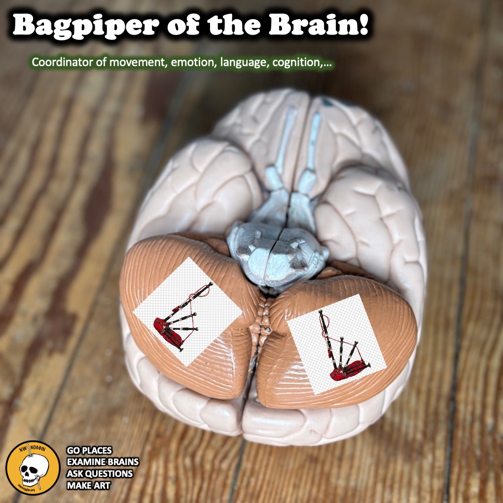
The brain’s bagpiper!
Growing up as a competitive Scottish Highland dancer, I had no clue the cerebellum was basically the bagpiper of the brain. In some ways I wasn’t learning the dances, but my cerebellum was!
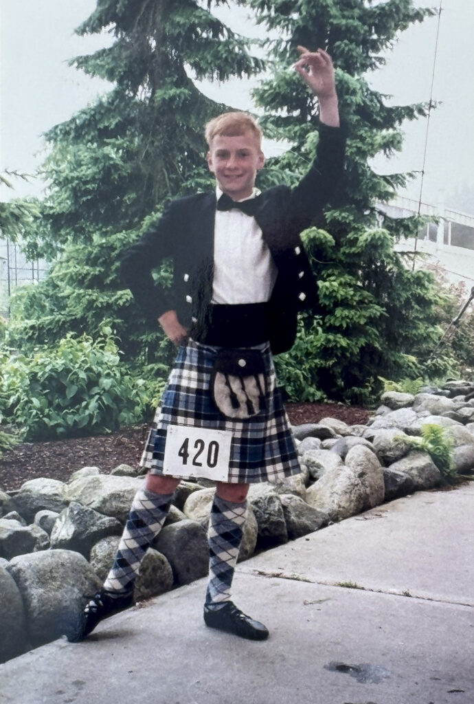
It kept me on beat and was also an important part of my brain being trained during the hours of practice I put in each week. Highland dancing requires a lot of precision, coordination and balance since you are jumping up and down on one foot a lot of the time and all three are the job of the cerebellum.
Fast forward to today, and it turns out I am still exercising my cerebellum for my new favorite hobby, skateboarding. Each time I get on my skateboard and try to learn a new trick, there I am once again developing greater precision in my movements, coordinating leg kicks with arm movements, and struggling to maintain balance as I work to stick the landing.
LEARN MORE: History of the Dances
LEARN MORE: Portland Highland Dance Association
LEARN MORE: Scotland Comes To The Pacific Northwest
LEARN MORE: The Dancing Brain: Structural and Functional Signatures of Expert Dance Training
LEARN MORE: Dancing and the Brain
What’s a cerebellum?
In every neuroscience course I have taken, the cerebellum was taught as being involved in motor functions like balance, posture, and movement coordination – and nothing more.
It has essentially been explained as a movement “supervisor,” observing and refining movements to make them smoother and more precise. This is accurate, but the cerebellum does so much more. Researchers have recently learned that it also plays a critical role in cognitive functions like decision making, language, emotions, and mood.
A whole world of cerebellar research has taken off over the last 30 years, and the more scientists examine this structure the more tasks it gets assigned. In the near future, watch as the connection between the cerebellum and cognition becomes better understood, especially its role in neurological conditions like schizophrenia and autism spectrum disorder (ASD). The cerebellum is an exciting area of research, and I am honored to have had the opportunity to learn more about it.
LEARN MORE: Consensus Paper: The Cerebellum’s Role in Movement and Cognition
LEARN MORE: The Role of the Cerebellum in Schizophrenia
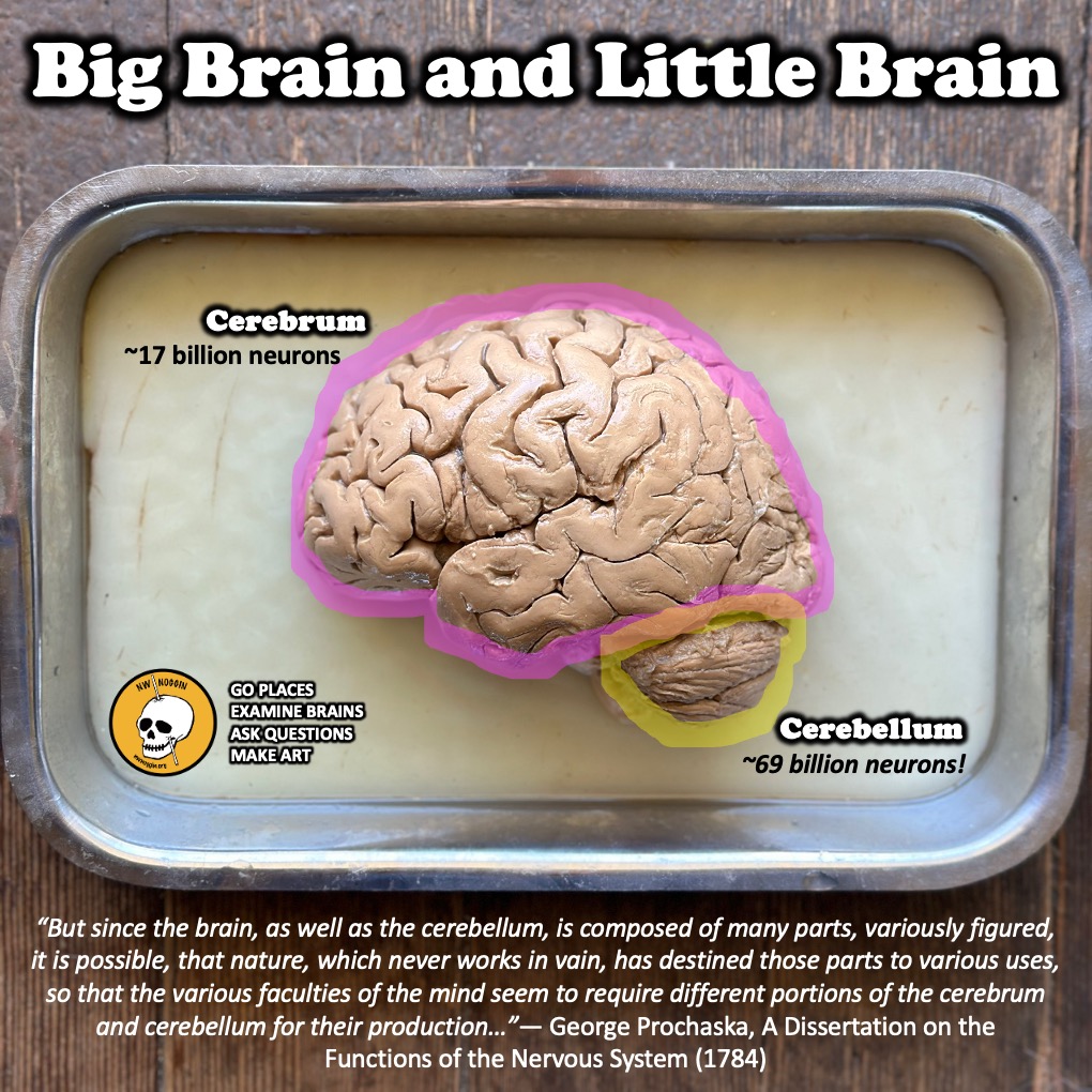
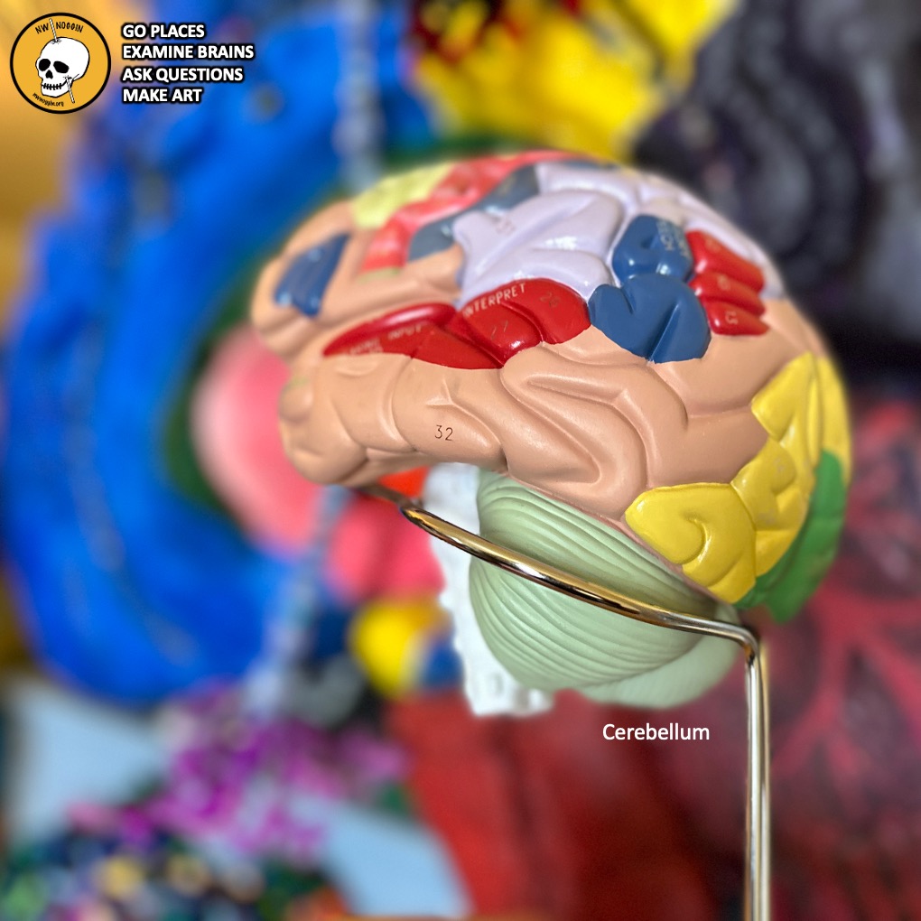
The CEREBRUM (big brain) is what most people picture when we think of the brain. It is the wrinkly pink mass that is responsible for everything from visual and auditory perception to our ability to make complex decisions and plan for the future. The CEREBELLUM (little brain) on the other hand is a small structure found attached to the big brain and is often overlooked.
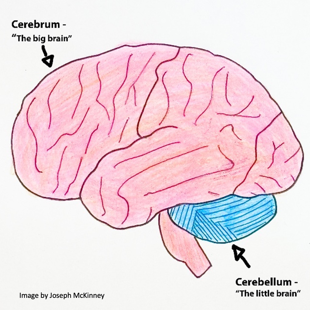
Cerebellum is a Latin word that literally means “little brain.”
It makes up only about 10% of the volume of the brain, but don’t let the name and size trick you because this “little” structure holds upwards of 80% of the billions of neurons that make up the whole brain, with some estimates suggesting somewhere around 69 billion neurons!
LEARN MORE: Soup for Brains!
LEARN MORE: The principles of physiology
Where is the Cerebellum?
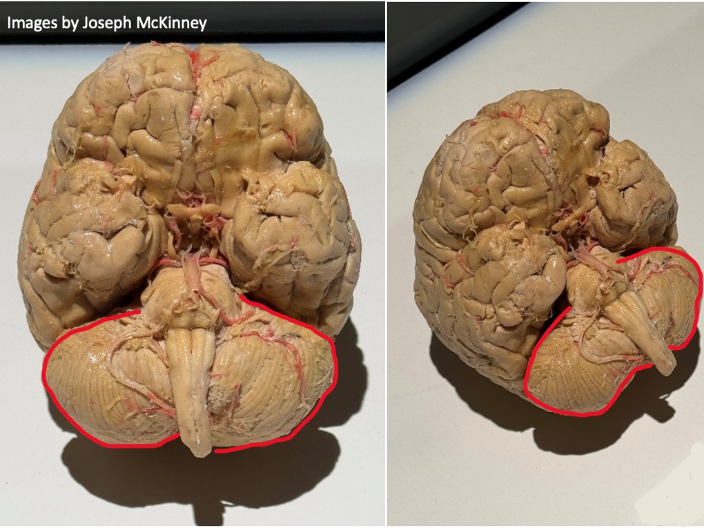
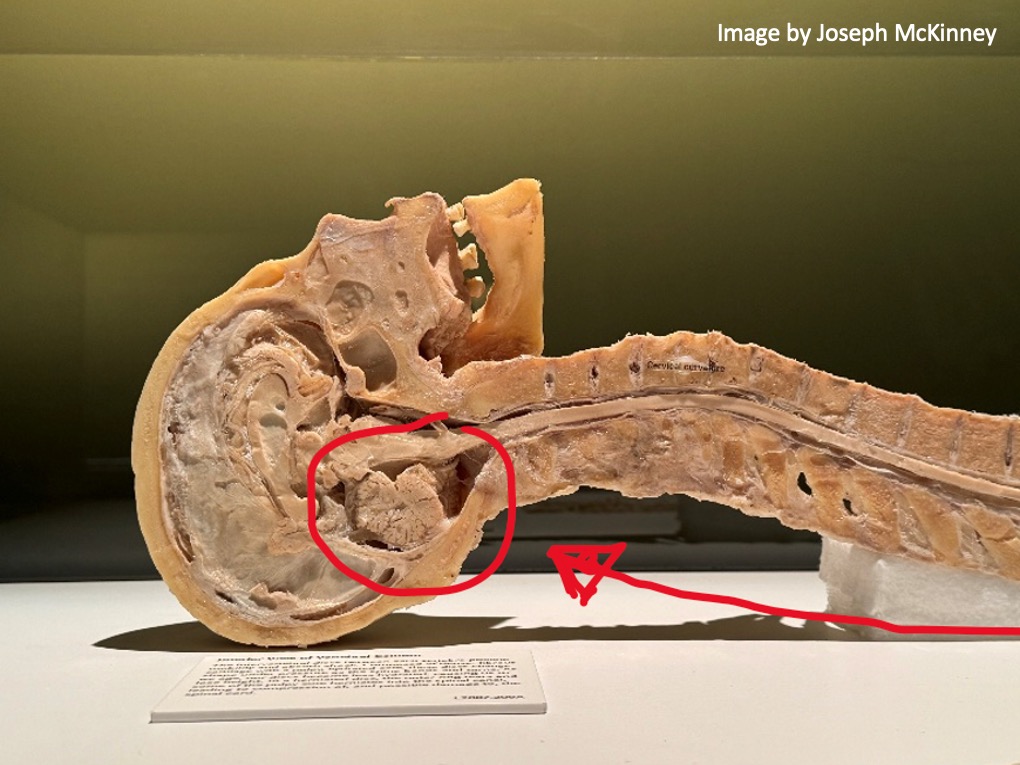
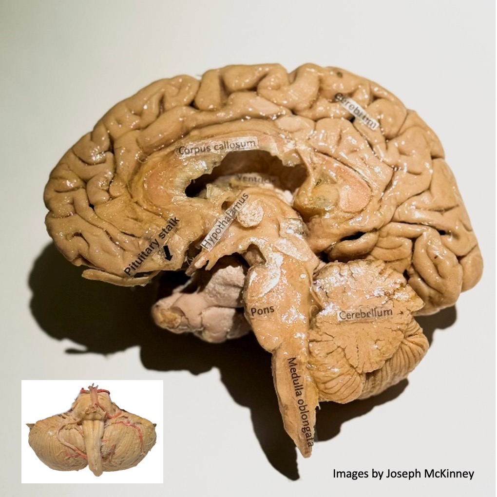
The cerebellum is found inferior to the cerebral hemispheres that make up the occipital lobe, and posterior to the brainstem structures called the pons and medulla oblongata. In normal words, that means it’s found attached to the back and bottom of the brain, right below the part of the brain that processes vision, and just behind the brainstem.
What does the cerebellum look like?
The more I looked at a real cerebellum during our outreach visits, the more I realized it really does look like a completely different structure than the rest of the brain, and that led me to the subtitle for this post, “The Big Brain’s Little Brain.”
The cerebellum is very tightly folded.
Another way to think of it is that it is really wrinkly, or highly striated (striped), with each wrinkle called a folia (Latin for “leaves”). The folia allow the cerebellum to have an incredibly large surface area, keeping lots of neurons packed into a very small space. Together with the large surface area, the fact that most of the cerebellar neurons are exceptionally tiny makes for its huge number of cells.
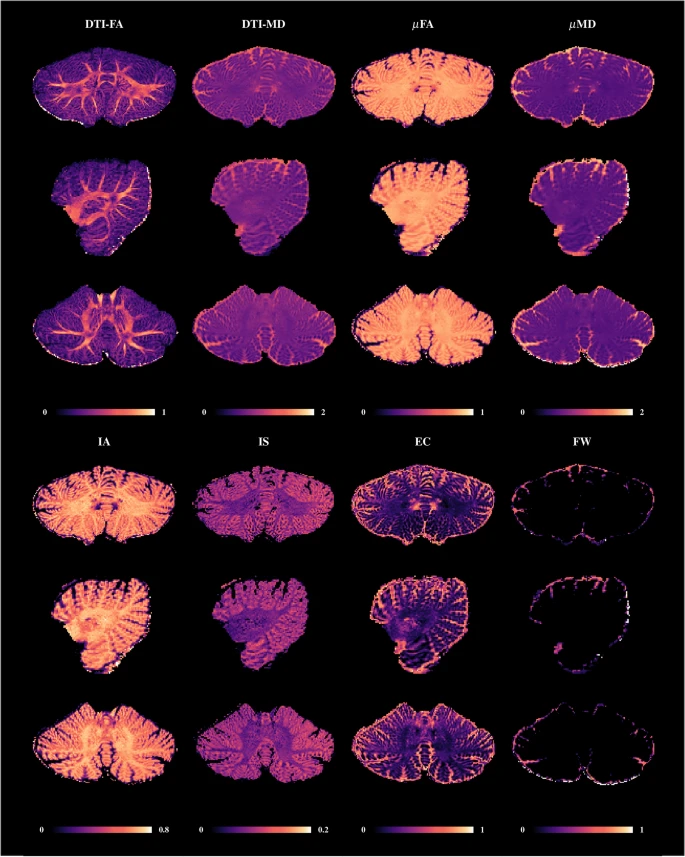
IMAGE SOURCE: A multimodal submillimeter MRI atlas of the human cerebellum
LEARN MORE: The Human Cerebellum: A Digital Anatomical Atlas at the Level of Individual Folia
LEARN MORE: Folia of human cerebellum: structure and variations
Some people think the cerebellum looks like cauliflower or a walnut, but I think it looks like a bundle of pasta because it appears striped, like it’s made of noodles. Don’t eat the cerebellum 👀
Made of cells
Every organ of every living thing is made of cells, and neurons are the type of cell that make up brains, including the cerebellum. Seeing and understanding the typical neuron is really helpful when it comes to understanding the specialized neurons that make up the cerebellum.
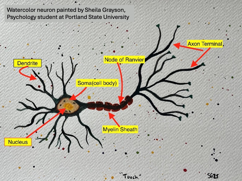
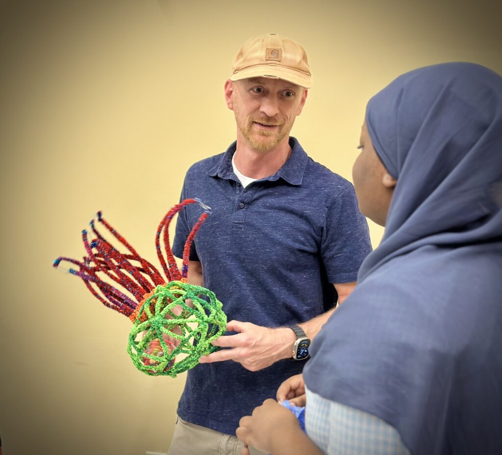
In Psychology 347 – Perception, an interdisciplinary course offered at Portland State University, students get to create their own neurons artistically to help understand their structure. Combining art with neuroscience is one way to help build more connections between different parts of the brain, and to make neuroscience even more fun.
LEARN MORE: STEAM Art Projects
LEARN MORE: Pipe Cleaner Brain Cells!
LEARN MORE: Portland State University and nonprofit NW Noggin
LEARN MORE: Golgi, Cajal and the Neuron Doctrine
The unique nature of the cellular structure of the cerebellum is one of the reasons I am so fascinated by it. Although the cerebellum itself is an extremely complex structure involved in many different aspects of life, the simple and repetitive layout of the cells shows just how efficient it is.
The key parts of neurons are the dendrites, which are kind of like the ears of a neuron sitting and waiting for a message, the soma (cell body) that holds the nucleus with all the DNA, the axon, which is the part of the neuron that sends a message along a neuron, and then the axon terminals, which release neurotransmitters (little chemical messages) onto the next neuron.

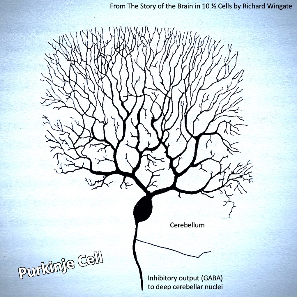
Purkinje Cells
A Purkinje cell is a special type of neuron found only in the cortex of the cerebellum.
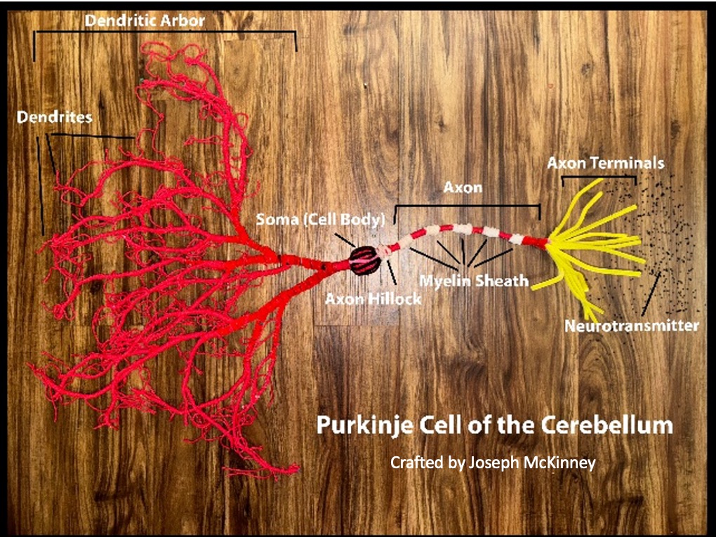
Their job is to take in tons of input, and to use that information to help coordinate movement with the muscles in the body via the deep cerebellar nuclei. They have a unique and extensive set of dendrites called the dendritic arbor, which allows them to connect with thousands of other neurons, to take in a ton of input at once, and to integrate that input into a single signal.
Purkinje cells are my favorite type of neuron because of their beautiful branching dendrites.
LEARN MORE: Histology, Purkinje Cells
Purkinje cells work by integrating their input into inhibitory signals sent to the deep cerebellar nuclei. The deep cerebellar nuclei (DCN) are the main sites where output leaves the cerebellum, and these outputs lead to the fine tuning of movements by making them smooth and coordinated.
Working in tandem with the basal ganglia, and almost every part of the cerebral cortex (the outer layer of the cerebrum), they help us do everything from riding a bike to speaking and threading needles.
Purkinje cells release a neurotransmitter called GABA, which has inhibitory roles in the nervous system, even calming anxiety. In motor functioning, GABA inhibits excessive or incorrect motor signals to prevent over-excitation. Keep these cells in mind for later when we talk about autism spectrum disorder.
LEARN MORE: Circuits within the Cerebellum
LEARN MORE: Integration of Purkinje Cell Inhibition by Cerebellar Nucleo-Olivary Neurons
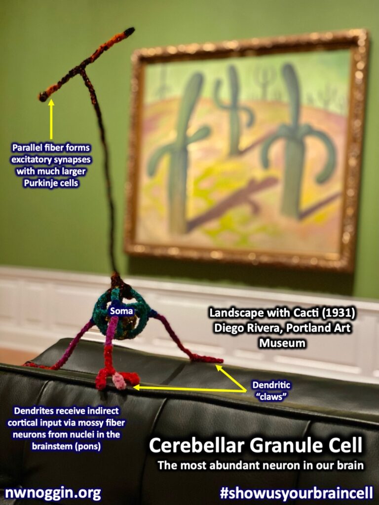
Granule cells
Cerebellar granule cells make up about 90-99% of the neurons in the cerebellum (depending on your source), and the cerebellum’s neurons account for about 80% of the neurons in the brain!
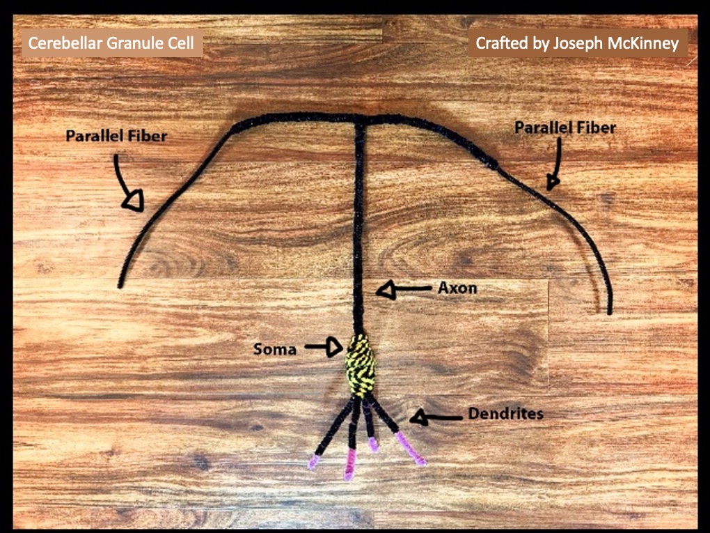
That means a HUGE proportion of our brain cells are cerebellar granule cells, which suggests they play an important role. The granule cells receive input from the rest of the brain through mossy fibers (a type of neuron) and Golgi cells (another type of neuron in the cerebellum) and then pass that input up along their axons to their parallel fibers. Parallel fibers are the two long branches splitting off the end of a granule cell’s axon in the molecular layer of the cerebellum.
Without the granule cells, Purkinje cells wouldn’t get very much information, and our movements would be jerky, rough, and uncoordinated. Imagine trying to grab a glass of water without being able to control the strength of your reach or grasp.
LEARN MORE: Origins, Development, and Compartmentation of the Granule Cells of the Cerebellum
LEARN MORE: The Significance of the Granular Layer of the Cerebellum
Cerebellar cortex is layered
The cerebellar cortex is composed of three main layers.

The molecular layer is the outermost layer where parallel fibers of granule cells will synapse with (pass a message to) the dendritic arbors of Purkinje cells. Below the molecular layer is the Purkinje layer, made up of cell bodies from the Purkinje cells. Then below the Purkinje layer is the granular layer. Can you guess what’s there? It’s the layer made up of the cell bodies of granule cells, and is where mossy fibers from the pontine nuclei, spinal cord, and the vestibulocochlear nerve synapse with the granule cells. These mossy fibers are carrying signals into the cerebellum, and they stimulate the granule cells, which will then send messages up their axons to their parallel fibers, which will signal the Purkinje cells.
LEARN MORE: Topsy Turvy: Functions of Climbing and Mossy Fibers in the Vestibulo-Cerebellum
LEARN MORE: Latest research on the anatomy and physiology of the cerebellum
Cerebellar anatomy
The cerebellum is made up of different parts, each with different functions, and it has bilateral symmetry. That means each different area of the cerebellum has a different job, and it is split into two hemispheres, with a left and a right side that are structurally similar.
Typically, the cerebellum is displayed as if it is one solid round structure, almost like a baseball. One thing I found to be interesting, however, is that it is really shaped more like a flat sheet or long oval that has been rolled up into itself, making it appear more rounded. This rolled up structure further increases the surface area of the cerebellum and helps allow for its incredible cell number.
NOTE: Drawing inspired by Thieme Atlas of Anatomy: Head and Neuroanatomy
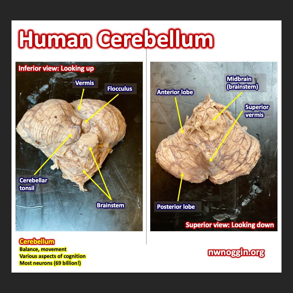
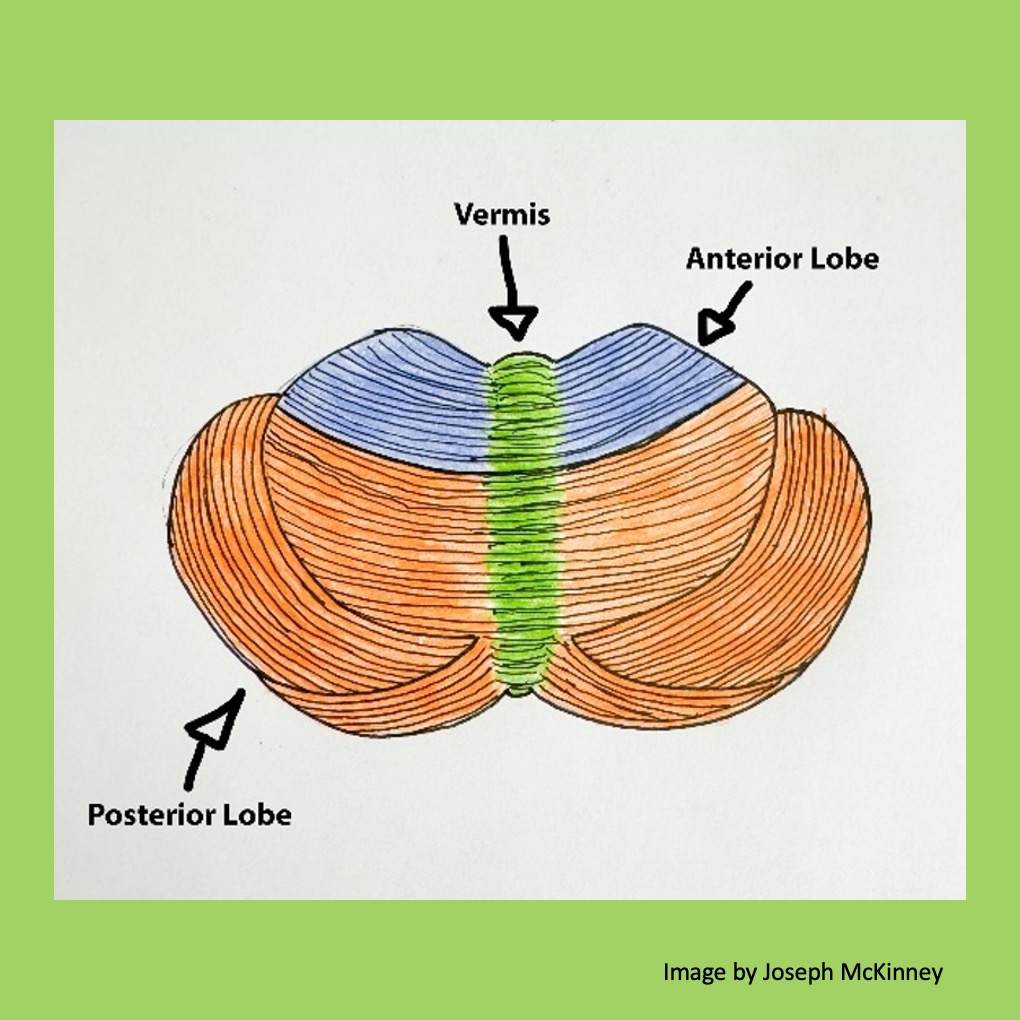
Anatomically the cerebellum is broken up into four main parts: the vermis, the anterior (front) lobe, the posterior (back) lobe, and the flocculonodular lobe. The image above shows the cerebellum in its rolled up form, the way it is found in the brain with the flocculonodular lobe and top of the vermis tucked into the structure, sort of like a rolled up potato bug.

This second image shows the cerebellum if it were to become unrolled, which would allow us to see the flocculonodular lobe. The cerebellum, like any anatomical structure, can also be referred to as having both a lateral (on the side, away from the middle) portion and a medial (towards the middle) portion.
What do these parts do?
In neuroanatomy, you often find the brain broken up into different parts in two different ways: one based on anatomy, so what part is where, and the other based on functionality, or what part does what job. The functional areas are composed of varying networks of the different anatomical areas described above.
Cerebrocerebellum
The cerebrocerebellum is like our little movement planning pal in the cerebellum.
It helps us perform all our movements in a smooth and coordinated fashion. The sides of both the front and back lobes (lateral portions) are where you find it. It is heavily involved in planning movements and motor learning.
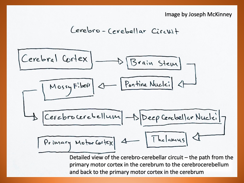
The cerebrocerebellum receives its inputs from the cerebral cortex and the pontine nuclei.
The pontine nuclei are a collection of cell bodies in the pons of the brainstem that will send a signal to the cerebrocerebellum, which then integrates the signal with other input and sends a refined movement plan to the thalamus. The thalamus is like the train station of the brain, where almost all information comes in and goes out. From here the signal is distributed to the motor cortex in the frontal lobes and the basal ganglia, two essential areas in the brain for executing movements.
Spinocerebellum
The spinocerebellum is kind of like the baby monitor of the brain – it keeps an eye on where our bodies are and what they’re doing.
It’s made up of the vermis and the middle (medial) portions of the front and back lobes of the cerebellum. The spinocerebellum receives tons of proprioceptive input, information about where the body is in space and what different parts of the body are doing.
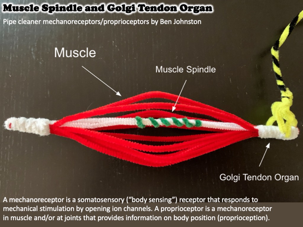
We have proprioceptive receptors all over our bodies in the muscles and joints, and these receptors continuously send positional and kinesthetic (movement) information to the spinocerebellum.
LEARN MORE: Flex Your Muscles … and Your Brain!

For a fun way to experience proprioception, close your eyes and touch your nose. You are able to do this because the cerebellum knows exactly where your face, arms, and hands are even without the eyes being involved.
LEARN MORE: Proprioception from a spinocerebellar perspective
Vestibulocerebellum
If the cerebrocerebellum is our built-in movement assistant, then the vestibulocerebellum is like our balance assistant. It helps us control our muscles to maintain balance and is also crucial for visual reflexes, helping us maintain our gaze on an object even when the head is moving.

The vestibulocerebellum is made up of the flocculonodular lobe, a little piece of the cerebellum that is tucked up inside of it. It receives input from the vestibular system (the system that controls balance) and sends outputs to the vestibular nuclei, a series of nuclei that are found in the pons and medulla oblongata of the brain stem.
To experience the vestibulocerebellum and its impact on our eyes, you can hold your finger up in front of your eyes and focus on your finger. If you move your head from side to side or up and down, you will see that you are able to maintain your gaze on your finger without any effort at all. This is the vestibulocerebellum in action!
Cerebellum and movement
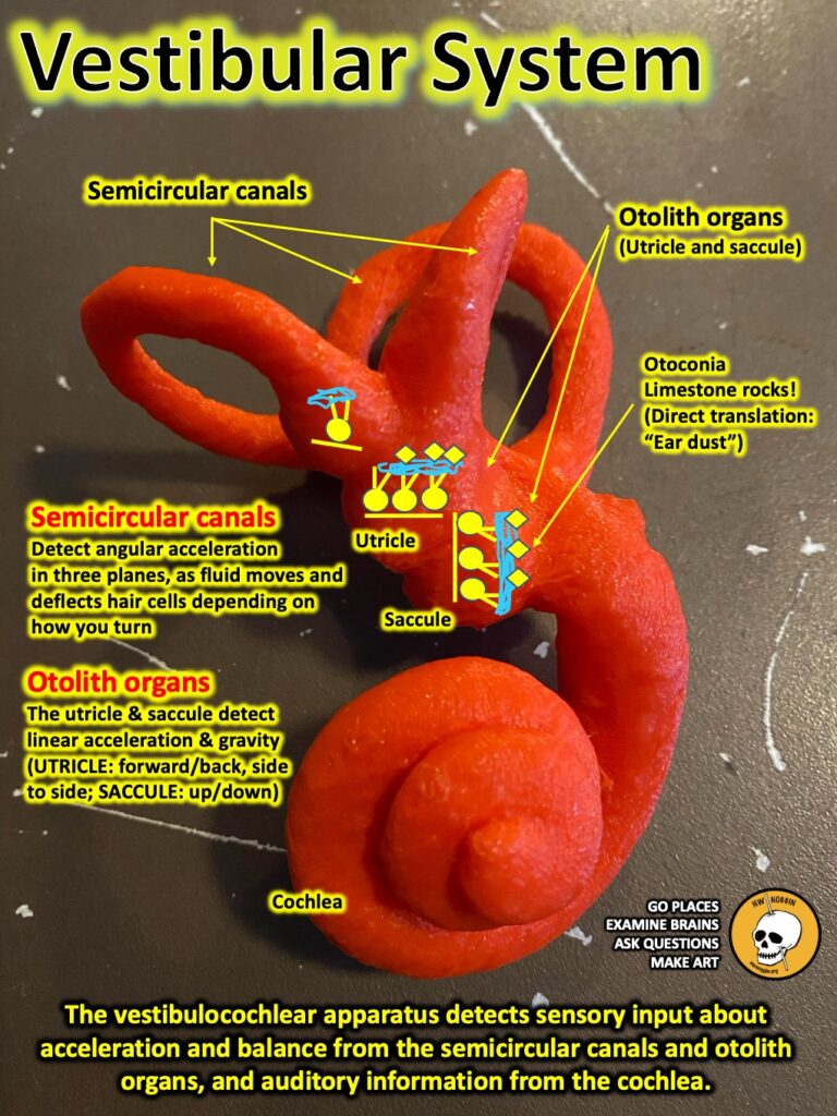
The major roles of the cerebellum are communication with our muscles and joints to facilitate the smoothing and coordination of movement and working with the vestibular system to maintain balance and posture. It constantly receives input from the muscles and joints in the body as well as the vestibular apparatus inside our inner ear to provide motor signals out to the body to accomplish these tasks. Imagine trying to stand on a horse if you have no idea of what your body is doing or no balance.
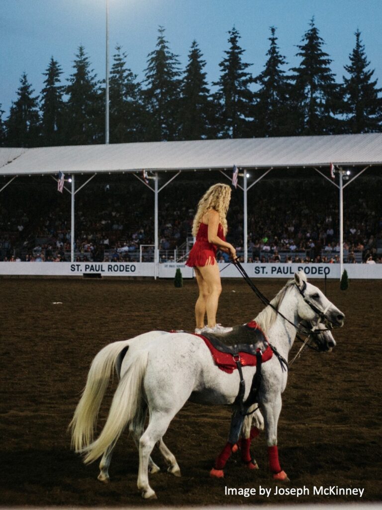
LEARN MORE: St. Paul Rodeo
Coordination, smoothing, and timing.
Say you decide to tie your shoe.
The motor cortex in the frontal lobe of the brain sends a motor plan for the planned movements to the pontine nuclei in the pons, where the message is then passed through the cerebellar peduncles (white matter cabling) to the cerebellum. At the same time, that motor plan is also sent to the muscles of the body to initiate the desired movements.
The cerebellum works really hard to collect information from our sensory systems via the Purkinje cells so it knows exactly what the body is doing and where it is millisecond by millisecond. This allows it to compare the motor plan to what is happening in real time.
If the cerebellum detects a mismatch between the desired movements and what is actually happening, it quickly computes the necessary changes to make the desired movements accurate.
Once the necessary changes are calculated, Purkinje cells send an inhibitory signal to the deep cerebellar nuclei, including the dentate nuclei, which then send corrective signals to our brainstem and spinal cord, and to our motor cortex via the thalamus. The motor cortex then sends a new signal down to the muscles of the body and the mismatch is fixed.
Through that process the cerebellum is able to support our movements by keeping them smooth (not jerky), to prevent over or undershooting a desired movement, and it keeps the timing of the activation of different muscles accurate. Imagine how hard it would be to speak if the muscles of articulation (mouth muscles, tongue) were activated in the wrong order. For those with cerebellar damage it is problematic.
Since we already learned how the vestibulo-cerebellar circuit works, now let’s see what it does. The purpose of the vestibular system is to detect head tilt or movement in different directions, and to send that information through the vestibulocochlear nerve (CNVIII) from the inner ear to the brainstem and then different areas of the brain. My focus is the pathway that leads to the cerebellum.
First, some kind of tilt is detected by the hair cells in the vestibular apparatus, and then the signal is passed to the vestibular nuclei in the pons and medulla of the brainstem. Once the signal arrives at the brainstem, it is passed through the cerebellar peduncles (lots of white matter axons) that branch off the pons to the flocculonodular lobe in the cerebellum, which integrates the sensory info and sends a motor response back to the vestibular nuclei to recruit the trunk and limb muscles. This motor signal will stimulate muscles that will allow us to stay upright.

IMAGE SOURCE: Cerebellum: Sensing how to balance
LEARN MORE: Physiology of the cerebellum
Cerebellum and cognition
Cognition is a term that encompasses many of the different things we associate with the brain, but simply put it is our ability to think, learn, and understand things happening in and around us. Because research has primarily focused on the motor functions of the cerebellum, its contributions to cognition are far less understood, which will be reflected in the shorter explanations below. However, newer research does indicate that the cerebellum also plays a crucial role in cognitive functioning.
LEARN MORE: The cerebellum and cognition
LEARN MORE: Neocortex–Cerebellum Circuits for Cognitive Processing
Language
Speech is a motor function that involves the use of lots of muscles to move parts of the larynx (voice box), tongue and mouth, so it should come as no surprise that the cerebellum is heavily involved in speech. However, more recently researchers have begun to understand that language, a cognitive function requiring coordination of multiple parts of the brain, also has cerebellar involvement.
There are two main ways this connection is researched: using imaging studies like fMRI where scientists take pictures that show what parts of the brain are active during different tasks, and through correlational studies that show connections between damage to the cerebellum and changes in language ability. For example, imaging studies make it clear that when we use and listen to language, the cerebellum is highly active. Correlational studies have also suggested that people with cerebellar damage due to stroke often show clear impairments to language – particularly in semantic knowledge and the ability to form connections between nouns and verbs. An example of semantic knowledge could be understanding that a cat or dog is a type of animal, and an example of the inability to connect nouns with verbs could be not understanding that a ball (noun) is something you throw (verb).
LEARN MORE: Cerebellar Contributions to Language in Typical and Atypical Development: A Review
LEARN MORE: Language and the Cerebellum: Structural Connectivity to the Eloquent Brain
Social Cognition
Social cognition can be thought of as how we understand other people’s intentions and emotions from their verbal and nonverbal cues. An example of this could be looking at a person’s facial expressions and understanding their emotional state.
There are very few studies that specifically focus on the correlation between cerebellar damage and changes in social cognition, but many whole brain fMRI studies have shown that the cerebellum is active during these tasks.
Because this area of research is so under-explored it is hard to draw strong conclusions, but it does suggest that the cerebellum plays a role in social cognition and is a great area of research to expand on – providing opportunities for young scientists like the ones we met during outreach visits.
LEARN MORE: Little brain, little minds: The big role of the cerebellum in social development
LEARN MORE: The Social Cerebellum: A Large-Scale Investigation of Functional and Structural Specificity and Connectivity
LEARN MORE: Consensus Paper: Cerebellum and Social Cognition
Affect
There are multiple areas of the brain that are associated with affect and emotional processing. These areas include the hypothalamus, different cortical and subcortical parts of the limbic system, as well as association areas in different lobes of cortex.
Historically these brain areas were thought to be relatively distinct from each other, but research has begun to reveal that they may form a large and coordinated brain network that uses the cerebellum as a central hub for emotional processing – perhaps like its involvement in movement where it helps to prevent exaggerated or uncoordinated responses.
LEARN MORE: The Cerebellar Role in Emotions at a Turning Point
LEARN MORE: The Cerebellum and Emotional Experience
LEARN MORE: Cerebellum and Emotion Processing
Clinical Implications
There are lots of clinical consequences from damage to the cerebellum. Loss of coordination and balance is referred to as ataxia. Damage might also cause problems with expressing yourself through and understanding language leading a person to feel trapped and disconnected from the world around them, or they could experience extreme mood swings or depression. Additionally, since the cerebellum is connected with so many other brain areas, dysfunction could contribute to conditions like autism spectrum disorder, schizophrenia, and depression.
The cerebellum is not just a fascinating area of research, but potentially an incredibly important one.
LEARN MORE: Mechanisms of cerebellar gait ataxia
LEARN MORE: Ignoring the cerebellum is hindering progress in neuroscience
The cerebellum and autism spectrum disorder (ASD)
Being that the cerebellum likely plays a role in social cognition, emotion, and affect, some researchers have begun to hypothesize that the cerebellum could be involved in autism spectrum disorder. More specifically, they think Purkinje cells may play a role. Although there is no definitive link, evidence for this hypothesis has come from different types of studies. In one study, researchers looked at post-mortem brains from people with ASD and found that some show a significantly decreased number of these cells. In another type of study, when certain genes that are commonly thought to be associated with ASD are turned off in the Purkinje cells of mice, they can begin to show similar behaviors to people with the disorder, like social impairment and repetitive behaviors. Although none of this research is definitive, and all of it needs to be expanded on, it could have very large clinical implications for treatments and coping strategies and is important to point out.
LEARN MORE: The Cerebellar Involvement in Autism Spectrum Disorders
LEARN MORE: Neuroanatomy of autism: what is the role of the cerebellum?
Maintaining my balance
Learning about the cerebellum has shown me there are lots of opportunities for budding neuroscience enthusiasts like me and the students I met with NWNoggin to contribute to this field in meaningful ways. I also learned that one of the things I am curious about is how all the different brain structures interact with each other to create our lived experience.
It has shown me how important neuroimaging and our ability to analyze brain data is and has been a push for me to commit to the Neuroimaging and Neuroinformatics program at the University of Southern California next fall, where I will learn to do brain scans using MRI and fMRI, and to build the software necessary to analyze the mountains of data they produce.
LEARN MORE: USC Master of Science in Neuroimaging and Informatics
Volunteering with NWNoggin has been a highlight of my time at Portland State University.
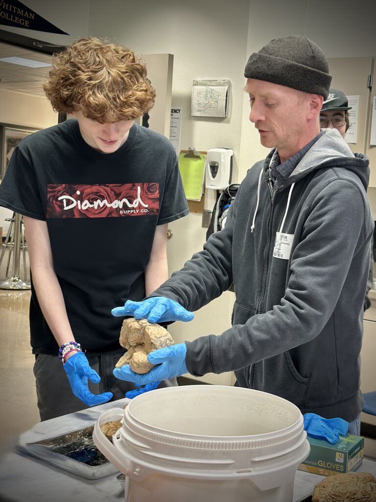
Connecting with community members showed me how much interest people have in the brain (it’s a lot!) and gave me the opportunity to hear the questions people really want answered. Their questions motivated me to understand neuroscience better so I could explain it in an approachable way and was what I needed to push through the “senioritis” that can come in a person’s final term of school. Lastly, I was able to hold real human brains! This is an experience I wish more people got to have as holding and thinking about the thing that thinks is amazing. The connection this experience gave me to a structure that is otherwise hidden and mysterious was more than just fun, it was life changing and I am forever grateful to NWNoggin for this experience.